

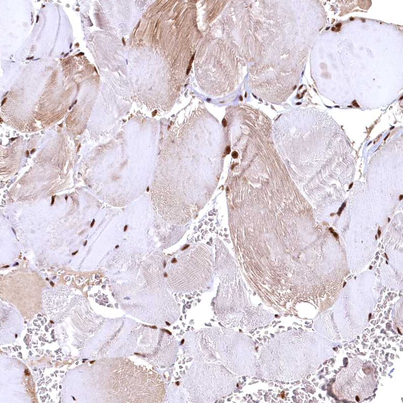
Immunohistochemical staining of human fallopian tube shows strong nuclear positivity in glandular cells.
 and RSRC2 over-expression lysate (Co-expressed with a C-terminal myc-DDK tag (~3.1 kDa) in mammalian HEK293T cells, LY411486).")
Immunohistochemical staining of human fallopian tube shows strong nuclear positivity in glandular cells.
Anti-RSRC2 Antibody
HPA040070
ApplicationsWestern Blot, ImmunoCytoChemistry, ImmunoHistoChemistry
Product group Antibodies
ReactivityHuman
TargetRSRC2
Overview
- SupplierAtlas Antibodies
- Product NameAnti-RSRC2 Antibody
- Delivery Days Customer4
- ApplicationsWestern Blot, ImmunoCytoChemistry, ImmunoHistoChemistry
- CertificationResearch Use Only
- ClonalityPolyclonal
- ConjugateUnconjugated
- Gene ID65117
- Target nameRSRC2
- Target descriptionarginine and serine rich coiled-coil 2
- Target synonymsarginine/serine-rich coiled-coil protein 2, arginine/serine-rich coiled-coil 2
- HostRabbit
- IsotypeIgG
- Protein IDQ7L4I2
- Protein NameArginine/serine-rich coiled-coil protein 2
- Scientific DescriptionRecombinant Protein Epitope Signature Tag (PrEST) antigen sequence
- ReactivityHuman
- Storage Instruction-20°C,2°C to 8°C
- UNSPSC41116161
Datasheet
MSDS
Related products

Product group Antibodies
Anti-RSRC2 Antibody Picoband(r)A13417-1-CARRIER-FREE
ApplicationsImmunoFluorescence, Western Blot, ELISA, ImmunoHistoChemistry
ReactivityHuman
TargetRSRC2
- SizePrice

Product group Antibodies

RSRC2 Antibody (aa8-37)LS-C158903
ApplicationsWestern Blot
ReactivityHuman
TargetRSRC2
- SizePrice

Product group Antibodies
Anti-RSRC2 AntibodyHPA048183
ApplicationsImmunoCytoChemistry
ReactivityHuman
TargetRSRC2
- SizePrice
Product group Antibodies
RSRC2 antibody, C-termGTX45867
ApplicationsWestern Blot, ImmunoHistoChemistry, ImmunoHistoChemistry Paraffin
ReactivityHuman
TargetRSRC2
- SizePrice
Product group Antibodies
Anti-RSRC2 (N-term) Antibody102-22878
ApplicationsWestern Blot
TargetRSRC2
- SizePrice
Product group Antibodies
RSRC2 Polyclonal AntibodyBS-6097R
ApplicationsImmunoFluorescence, Western Blot, ELISA, ImmunoCytoChemistry, ImmunoHistoChemistry, ImmunoHistoChemistry Frozen, ImmunoHistoChemistry Paraffin
ReactivityBovine, Canine, Chicken, Equine, Human, Mouse, Porcine, Rat
TargetRSRC2
- SizePrice