


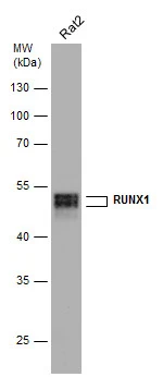
Figure 1. Western blot analysis of RUNX1 using anti-RUNX1 antibody (PB9157). Electrophoresis was performed on a 5-20% SDS-PAGE gel at 70V (Stacking gel) / 90V (Resolving gel) for 2-3 hours. The sample well of each lane was loaded with 50ug of sample under reducing conditions. Lane 1: human HL-60 whole cell lysates, Lane 2: rat thymus tissue lysates, Lane 3: mouse thymus tissue lysates, After Electrophoresis, proteins were transferred to a Nitrocellulose membrane at 150mA for 50-90 minutes. Blocked the membrane with 5% Non-fat Milk/ TBS for 1.5 hour at RT. The membrane was incubated with rabbit anti-RUNX1 antigen affinity purified polyclonal antibody (Catalog # PB9157) at 0.5 microg/mL overnight at 4°C, then washed with TBS-0.1%Tween 3 times with 5 minutes each and probed with a goat anti-rabbit IgG-HRP secondary antibody at a dilution of 1:10000 for 1.5 hour at RT. The signal is developed using an Enhanced Chemiluminescent detection (ECL) kit (Catalog # EK1002) with Tanon 5200 system. A specific band was detected for RUNX1 at approximately 55KD. The expected band size for RUNX1 is at 49KD.
. RUNX1 was detected in paraffin-embedded section of rat thymus tissue. Heat mediated antigen retrieval was performed in citrate buffer (pH6, epitope retrieval solution) for 20 mins. The tissue section was blocked with 10% goat serum. The tissue section was then incubated with 1microg/ml rabbit anti-RUNX1 Antibody (PB9157) overnight at 4°C. Biotinylated goat anti-rabbit IgG was used as secondary antibody and incubated for 30 minutes at 37°C. The tissue section was developed using Strepavidin-Biotin-Complex (SABC)(Catalog # SA1022) with DAB as the chromogen.")
. RUNX2 was detected in paraffin-embedded section of human mammary cancer tissue. Heat mediated antigen retrieval was performed in citrate buffer (pH6, epitope retrieval solution) for 20 mins. The tissue section was blocked with 10% goat serum. The tissue section was then incubated with 1microg/ml rabbit anti-RUNX2 Antibody (PB9157) overnight at 4°C. Biotinylated goat anti-rabbit IgG was used as secondary antibody and incubated for 30 minutes at 37°C. The tissue section was developed using Strepavidin-Biotin-Complex (SABC)(Catalog # SA1022) with DAB as the chromogen.")
. RUNX3 was detected in paraffin-embedded section of mouse intestine tissue. Heat mediated antigen retrieval was performed in citrate buffer (pH6, epitope retrieval solution) for 20 mins. The tissue section was blocked with 10% goat serum. The tissue section was then incubated with 1microg/ml rabbit anti-RUNX3 Antibody (PB9157) overnight at 4°C. Biotinylated goat anti-rabbit IgG was used as secondary antibody and incubated for 30 minutes at 37°C. The tissue section was developed using Strepavidin-Biotin-Complex (SABC)(Catalog # SA1022) with DAB as the chromogen.")
. RUNX3 was detected in paraffin-embedded section of rat intestine tissue. Heat mediated antigen retrieval was performed in citrate buffer (pH6, epitope retrieval solution) for 20 mins. The tissue section was blocked with 10% goat serum. The tissue section was then incubated with 1microg/ml rabbit anti-RUNX3 Antibody (PB9157) overnight at 4°C. Biotinylated goat anti-rabbit IgG was used as secondary antibody and incubated for 30 minutes at 37°C. The tissue section was developed using Strepavidin-Biotin-Complex (SABC)(Catalog # SA1022) with DAB as the chromogen.")
. RUNX3 was detected in frozen section of rat spleen tissue. The tissue section was blocked with 10% goat serum. The tissue section was then incubated with 1microg/ml rabbit anti-RUNX3 Antibody (PB9157) overnight at 4°C. Biotinylated goat anti-rabbit IgG was used as secondary antibody and incubated for 30 minutes at 37°C. The tissue section was developed using Strepavidin-Biotin-Complex (SABC)(Catalog # SA1022) with DAB as the chromogen.")
Figure 1. Western blot analysis of RUNX1 using anti-RUNX1 antibody (PB9157). Electrophoresis was performed on a 5-20% SDS-PAGE gel at 70V (Stacking gel) / 90V (Resolving gel) for 2-3 hours. The sample well of each lane was loaded with 50ug of sample under reducing conditions. Lane 1: human HL-60 whole cell lysates, Lane 2: rat thymus tissue lysates, Lane 3: mouse thymus tissue lysates, After Electrophoresis, proteins were transferred to a Nitrocellulose membrane at 150mA for 50-90 minutes. Blocked the membrane with 5% Non-fat Milk/ TBS for 1.5 hour at RT. The membrane was incubated with rabbit anti-RUNX1 antigen affinity purified polyclonal antibody (Catalog # PB9157) at 0.5 microg/mL overnight at 4°C, then washed with TBS-0.1%Tween 3 times with 5 minutes each and probed with a goat anti-rabbit IgG-HRP secondary antibody at a dilution of 1:10000 for 1.5 hour at RT. The signal is developed using an Enhanced Chemiluminescent detection (ECL) kit (Catalog # EK1002) with Tanon 5200 system. A specific band was detected for RUNX1 at approximately 55KD. The expected band size for RUNX1 is at 49KD.
Anti-RUNX1/AML1 Antibody Picoband(r)
PB9157-CARRIER-FREE
ApplicationsWestern Blot, ImmunoHistoChemistry, ImmunoHistoChemistry Frozen
Product group Antibodies
ReactivityHamster, Human, Mouse, Rat
TargetRUNX1
Overview
- SupplierBoster Bio
- Product NameAnti-RUNX1/AML1 Antibody Picoband(r)
- Delivery Days Customer9
- Application Supplier NoteWB: The detection limit for RUNX1 is approximately 0.25ng/lane under reducing conditions. Tested Species: In-house tested species with positive results. By Heat: Boiling the paraffin sections in 10mM citrate buffer, pH6.0, for 20mins is required for the staining of formalin/paraffin sections. Other applications have not been tested. Optimal dilutions should be determined by end users.
- ApplicationsWestern Blot, ImmunoHistoChemistry, ImmunoHistoChemistry Frozen
- CertificationResearch Use Only
- ClonalityPolyclonal
- Concentration500 ug/ml
- Gene ID861
- Target nameRUNX1
- Target descriptionRUNX family transcription factor 1
- Target synonymsAML1, AML1-EVI-1, AMLCR1, CBF2alpha, CBFA2, EVI-1, PEBP2aB, PEBP2alpha, runt-related transcription factor 1, AML1-ETO fusion, AML1-ETO fusion protein, AML1-EVI-1 fusion protein, PEA2-alpha B, PEBP2-alpha B, SL3-3 enhancer factor 1 alpha B subunit, SL3/AKV core-binding factor alpha B subunit, acute myeloid leukemia 1 protein, core-binding factor, runt domain, alpha subunit 2, mutant RUNX1, oncogene AML-1, polyomavirus enhancer-binding protein 2 alpha B subunit, runt related transcription factor 1
- HostRabbit
- IsotypeIgG
- Protein IDQ01196
- Protein NameRunt-related transcription factor 1
- Scientific DescriptionBoster Bio Anti-RUNX1/AML1 Antibody Picoband® catalog # PB9157. Tested in IHC, IHC-F, WB applications. This antibody reacts with Human, Mouse, Rat. The brand Picoband indicates this is a premium antibody that guarantees superior quality, high affinity, and strong signals with minimal background in Western blot applications. Only our best-performing antibodies are designated as Picoband, ensuring unmatched performance.
- ReactivityHamster, Human, Mouse, Rat
- Storage Instruction-20°C,2°C to 8°C
- UNSPSC12352203
Related products
Product group Antibodies

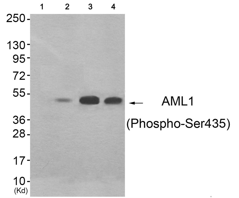
AML1 (Phospho-Ser435) AntibodyABX012668
ApplicationsWestern Blot, ELISA
- SizePrice
Product group Antibodies
Anti-RUNX1 Antibody144-02055
ApplicationsImmunoFluorescence, Western Blot, ImmunoHistoChemistry
ReactivityHuman, Mouse, Rat
TargetRUNX1
- SizePrice
Product group Antibodies
ApplicationsWestern Blot
ReactivityHuman
- SizePrice
Product group Antibodies
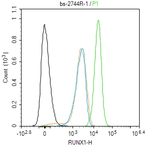
RUNX1 Polyclonal Antibodybs-2744R
ApplicationsFlow Cytometry, Western Blot, ELISA, ImmunoHistoChemistry, ImmunoHistoChemistry Paraffin
ReactivityBovine, Canine, Equine, Human, Mouse, Rabbit, Rat
TargetRUNX1
- SizePrice
Product group Antibodies
RUNX1 AntibodyCSB-PA010571
ApplicationsImmunoFluorescence, Western Blot, ELISA, ImmunoHistoChemistry
ReactivityHuman, Mouse, Rat
TargetRUNX1
- SizePrice
Product group Antibodies
RUNX1 Polyclonal AntibodyCAC14795
ApplicationsImmunoFluorescence, Western Blot, ELISA
TargetRUNX1
- SizePrice
Product group Antibodies

AML1 / RUNX1 Antibody (phospho-Ser276)LS-C358858
ApplicationsWestern Blot, ImmunoHistoChemistry, ImmunoHistoChemistry Paraffin
ReactivityChicken, Human, Mouse, Porcine, Rat
TargetRUNX1
- SizePrice

Product group Antibodies
Anti-RUNX1 AntibodyHPA004176
ApplicationsImmunoCytoChemistry, ImmunoHistoChemistry
ReactivityHuman
TargetRUNX1
- SizePrice
Product group Antibodies
RUNX1 antibodyGTX129100
ApplicationsImmunoFluorescence, Western Blot, ImmunoCytoChemistry, ImmunoHistoChemistry
ReactivityHuman, Mouse, Rat
TargetRUNX1
- SizePrice