


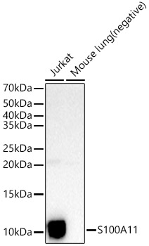
Figure 1. Western blot analysis of S100A11 using anti-S100A11 antibody (PB9818). Electrophoresis was performed on a 5-20% SDS-PAGE gel at 70V (Stacking gel) / 90V (Resolving gel) for 2-3 hours. The sample well of each lane was loaded with 40 ug of sample under reducing conditions. Lane 1: HELA Whole Cell Lysate. After electrophoresis, proteins were transferred to a nitrocellulose membrane at 150 mA for 50-90 minutes. Blocked the membrane with 5% non-fat milk/TBS for 1.5 hour at RT. The membrane was incubated with rabbit anti-S100A11 antigen affinity purified polyclonal antibody (Catalog # PB9818) at 0.5 microg/mL overnight at 4°C, then washed with TBS-0.1%Tween 3 times with 5 minutes each and probed with a goat anti-rabbit IgG-HRP secondary antibody at a dilution of 1:5000 for 1.5 hour at RT. The signal is developed using an Enhanced Chemiluminescent detection (ECL) kit (Catalog # EK1002) with Tanon 5200 system. A specific band was detected for S100A11 at approximately 15 kDa. The expected band size for S100A11 is at 15 kDa.
Figure 1. Western blot analysis of S100A11 using anti-S100A11 antibody (PB9818). Electrophoresis was performed on a 5-20% SDS-PAGE gel at 70V (Stacking gel) / 90V (Resolving gel) for 2-3 hours. The sample well of each lane was loaded with 40 ug of sample under reducing conditions. Lane 1: HELA Whole Cell Lysate. After electrophoresis, proteins were transferred to a nitrocellulose membrane at 150 mA for 50-90 minutes. Blocked the membrane with 5% non-fat milk/TBS for 1.5 hour at RT. The membrane was incubated with rabbit anti-S100A11 antigen affinity purified polyclonal antibody (Catalog # PB9818) at 0.5 microg/mL overnight at 4°C, then washed with TBS-0.1%Tween 3 times with 5 minutes each and probed with a goat anti-rabbit IgG-HRP secondary antibody at a dilution of 1:5000 for 1.5 hour at RT. The signal is developed using an Enhanced Chemiluminescent detection (ECL) kit (Catalog # EK1002) with Tanon 5200 system. A specific band was detected for S100A11 at approximately 15 kDa. The expected band size for S100A11 is at 15 kDa.
Anti-S100A11 Antibody Picoband(r)
PB9818-CARRIER-FREE
ApplicationsWestern Blot
Product group Antibodies
ReactivityHuman
TargetS100A11
Overview
- SupplierBoster Bio
- Product NameAnti-S100A11 Antibody Picoband(r)
- Delivery Days Customer9
- Application Supplier NoteTested Species: In-house tested species with positive results. Other applications have not been tested. Optimal dilutions should be determined by end users.
- ApplicationsWestern Blot
- CertificationResearch Use Only
- ClonalityPolyclonal
- Concentration500 ug/ml
- Gene ID6282
- Target nameS100A11
- Target descriptionS100 calcium binding protein A11
- Target synonymsHEL-S-43, MLN70, S100C, protein S100-A11, MLN 70, calgizzarin, epididymis secretory protein Li 43, metastatic lymph node gene 70 protein, protein S100-C
- HostRabbit
- IsotypeIgG
- Protein IDP31949
- Protein NameProtein S100-A11
- Scientific DescriptionBoster Bio Anti-S100A11 Antibody Picoband® catalog # PB9818. Tested in WB applications. This antibody reacts with Human. The brand Picoband indicates this is a premium antibody that guarantees superior quality, high affinity, and strong signals with minimal background in Western blot applications. Only our best-performing antibodies are designated as Picoband, ensuring unmatched performance.
- ReactivityHuman
- Storage Instruction-20°C,2°C to 8°C
- UNSPSC12352203
Related products
Product group Antibodies
Anti-S100A11 AntibodyA306920
ApplicationsImmunoFluorescence, Western Blot, ImmunoCytoChemistry
ReactivityHuman, Mouse
- SizePrice
Product group Antibodies
Anti-S100A11 Antibody130-10933-200
ApplicationsELISA
ReactivityHuman
TargetS100A11
- SizePrice
Product group Antibodies

Calgizzarin / S100A11 AntibodyLS-C831494
ApplicationsImmunoHistoChemistry
ReactivityHuman, Mouse
TargetS100A11
- SizePrice
Product group Antibodies
S100A11 Polyclonal AntibodyBS-55189R
ApplicationsImmunoFluorescence, Western Blot, ImmunoCytoChemistry
ReactivityHuman
TargetS100A11
- SizePrice
Product group Antibodies
ApplicationsWestern Blot, ELISA, ImmunoCytoChemistry, ImmunoHistoChemistry, ImmunoHistoChemistry Frozen, ImmunoHistoChemistry Paraffin
ReactivityMouse, Rat
TargetS100A11
- SizePrice
Product group Antibodies
S100A11 AntibodyCSB-PA020624HA01HU
ApplicationsELISA, ImmunoHistoChemistry
ReactivityHuman
TargetS100A11
- SizePrice
Product group Antibodies
S100A11 antibodyGTX100698
ApplicationsImmunoFluorescence, Western Blot, ImmunoCytoChemistry, ImmunoHistoChemistry, ImmunoHistoChemistry Paraffin
ReactivityHuman
TargetS100A11
- SizePrice

Product group Antibodies
Anti-S100A11 AntibodyHPA042745
ApplicationsImmunoHistoChemistry
ReactivityHuman
TargetS100A11
- SizePrice
Product group Antibodies
ApplicationsImmunoFluorescence, ImmunoPrecipitation, Western Blot, ELISA, ImmunoCytoChemistry
ReactivityHuman
TargetS100A11
- SizePrice