


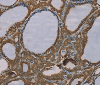
Immunohistochemical staining of human urinary bladder shows moderate cytoplasmic and nuclear positivity in urothelial cells.

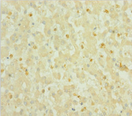
Immunohistochemical staining of human urinary bladder shows moderate cytoplasmic and nuclear positivity in urothelial cells.
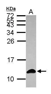
Anti-S100A6 Antibody
HPA007575
ApplicationsWestern Blot, ImmunoCytoChemistry, ImmunoHistoChemistry
Product group Antibodies
ReactivityHuman, Mouse, Rat
TargetS100A6
Overview
- SupplierAtlas Antibodies
- Product NameAnti-S100A6 Antibody
- Delivery Days Customer4
- ApplicationsWestern Blot, ImmunoCytoChemistry, ImmunoHistoChemistry
- CertificationResearch Use Only
- ClonalityPolyclonal
- ConjugateUnconjugated
- Gene ID6277
- Target nameS100A6
- Target descriptionS100 calcium binding protein A6
- Target synonyms2A9, 5B10, CABP, CACY, PRA, S10A6, protein S100-A6, MLN 4, calcyclin, growth factor-inducible protein 2A9, prolactin receptor-associated protein
- HostRabbit
- IsotypeIgG
- Protein IDP06703
- Protein NameProtein S100-A6
- Scientific DescriptionRecombinant Protein Epitope Signature Tag (PrEST) antigen sequence
- ReactivityHuman, Mouse, Rat
- Storage Instruction-20°C,2°C to 8°C
- UNSPSC41116161
Datasheet
MSDS
Related products
Product group Antibodies
Anti-S100A6 AntibodyA45600
ApplicationsImmunoHistoChemistry
ReactivityHuman
- SizePrice
Product group Antibodies
Anti-S100A6 Antibody144-04104
ApplicationsWestern Blot
ReactivityHuman, Mouse
TargetS100A6
- SizePrice
Product group Antibodies

S100A6 / Calcyclin AntibodyLS-C831548
ApplicationsImmunoHistoChemistry
ReactivityHuman, Rat
TargetS100A6
- SizePrice
Product group Antibodies
S100A6 Recombinant Antibody, AbBy Fluor-350 ConjugatedBSM-61393R-BF350
ApplicationsFlow Cytometry, ImmunoFluorescence, Western Blot
ReactivityHuman, Mouse, Rat
TargetS100A6
- SizePrice
Product group Antibodies
S100A6 AntibodyCSB-PA020634HA01HU
ApplicationsELISA, ImmunoHistoChemistry
ReactivityHuman
TargetS100A6
- SizePrice
Product group Antibodies
ApplicationsImmunoPrecipitation, Western Blot, ImmunoCytoChemistry, ImmunoHistoChemistry
ReactivityMouse, Rat
TargetS100A6
- SizePrice
Product group Antibodies
S100A6 antibodyGTX121460
ApplicationsImmunoFluorescence, Western Blot, ImmunoCytoChemistry
ReactivityHuman
TargetS100A6
- SizePrice

Product group Antibodies
Anti-S100 alpha 6/S100A6 Antibody Picoband(r)PB9676-CARRIER-FREE
ApplicationsFlow Cytometry, ImmunoFluorescence, Western Blot, ImmunoCytoChemistry, ImmunoHistoChemistry
ReactivityHuman, Mouse, Rat
TargetS100A6
- SizePrice