


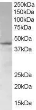
Figure 1. Western blot analysis of SAMSN1 using anti-SAMSN1 antibody (A08977-2). Electrophoresis was performed on a 5-20% SDS-PAGE gel at 70V (Stacking gel) / 90V (Resolving gel) for 2-3 hours. The sample well of each lane was loaded with 30 ug of sample under reducing conditions. Lane 1: human K562 whole cell lysates, Lane 2: human HEL whole cell lysates. After electrophoresis, proteins were transferred to a nitrocellulose membrane at 150 mA for 50-90 minutes. Blocked the membrane with 5% non-fat milk/TBS for 1.5 hour at RT. The membrane was incubated with rabbit anti-SAMSN1 antigen affinity purified polyclonal antibody (Catalog # A08977-2) at 0.5 microg/mL overnight at 4°C, then washed with TBS-0.1%Tween 3 times with 5 minutes each and probed with a goat anti-rabbit IgG-HRP secondary antibody at a dilution of 1:5000 for 1.5 hour at RT. The signal is developed using an Enhanced Chemiluminescent detection (ECL) kit (Catalog # EK1002) with Tanon 5200 system. A specific band was detected for SAMSN1 at approximately 50 kDa. The expected band size for SAMSN1 is at 42 kDa.
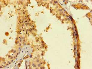
. SAMSN1 was detected in a paraffin-embedded section of human tonsil tissue. Heat mediated antigen retrieval was performed in EDTA buffer (pH 8.0, epitope retrieval solution). The tissue section was blocked with 10% goat serum. The tissue section was then incubated with 2 microg/ml rabbit anti-SAMSN1 Antibody (A08977-2) overnight at 4°C. Peroxidase Conjugated Goat Anti-rabbit IgG was used as secondary antibody and incubated for 30 minutes at 37°C. The tissue section was developed using HRP Conjugated Rabbit IgG Super Vision Assay Kit (Catalog # SV0002) with DAB as the chromogen.")
. SAMSN1 was detected in a paraffin-embedded section of mouse brain tissue. Heat mediated antigen retrieval was performed in EDTA buffer (pH 8.0, epitope retrieval solution). The tissue section was blocked with 10% goat serum. The tissue section was then incubated with 2 microg/ml rabbit anti-SAMSN1 Antibody (A08977-2) overnight at 4°C. Peroxidase Conjugated Goat Anti-rabbit IgG was used as secondary antibody and incubated for 30 minutes at 37°C. The tissue section was developed using HRP Conjugated Rabbit IgG Super Vision Assay Kit (Catalog # SV0002) with DAB as the chromogen.")
. SAMSN1 was detected in a paraffin-embedded section of rat brain tissue. Heat mediated antigen retrieval was performed in EDTA buffer (pH 8.0, epitope retrieval solution). The tissue section was blocked with 10% goat serum. The tissue section was then incubated with 2 microg/ml rabbit anti-SAMSN1 Antibody (A08977-2) overnight at 4°C. Peroxidase Conjugated Goat Anti-rabbit IgG was used as secondary antibody and incubated for 30 minutes at 37°C. The tissue section was developed using HRP Conjugated Rabbit IgG Super Vision Assay Kit (Catalog # SV0002) with DAB as the chromogen.")
. SAMSN1 was detected in an immunocytochemical section of HEL cells. Enzyme antigen retrieval was performed using IHC enzyme antigen retrieval reagent (AR0022) for 15 mins. The cells were blocked with 10% goat serum. And then incubated with 5 microg/mL rabbit anti-SAMSN1 Antibody (A08977-2) overnight at 4°C. DyLight488 Conjugated Goat Anti-Rabbit IgG (BA1127) was used as secondary antibody at 1:500 dilution and incubated for 30 minutes at 37°C. The section was counterstained with DAPI. Visualize using a fluorescence microscope and filter sets appropriate for the label used.")
. Overlay histogram showing K562 cells stained with A08977-2 (Blue line). To facilitate intracellular staining, cells were fixed with 4% paraformaldehyde and permeabilized with permeabilization buffer. The cells were blocked with 10% normal goat serum. And then incubated with rabbit anti-SAMSN1 Antibody (A08977-2, 1 microg/1x106 cells) for 30 min at 20°C. DyLight®488 conjugated goat anti-rabbit IgG (BA1127, 5-10 microg/1x106 cells) was used as secondary antibody for 30 minutes at 20°C. Isotype control antibody (Green line) was rabbit IgG (1 microg/1x106) used under the same conditions. Unlabelled sample (Red line) was also used as a control.")
Figure 1. Western blot analysis of SAMSN1 using anti-SAMSN1 antibody (A08977-2). Electrophoresis was performed on a 5-20% SDS-PAGE gel at 70V (Stacking gel) / 90V (Resolving gel) for 2-3 hours. The sample well of each lane was loaded with 30 ug of sample under reducing conditions. Lane 1: human K562 whole cell lysates, Lane 2: human HEL whole cell lysates. After electrophoresis, proteins were transferred to a nitrocellulose membrane at 150 mA for 50-90 minutes. Blocked the membrane with 5% non-fat milk/TBS for 1.5 hour at RT. The membrane was incubated with rabbit anti-SAMSN1 antigen affinity purified polyclonal antibody (Catalog # A08977-2) at 0.5 microg/mL overnight at 4°C, then washed with TBS-0.1%Tween 3 times with 5 minutes each and probed with a goat anti-rabbit IgG-HRP secondary antibody at a dilution of 1:5000 for 1.5 hour at RT. The signal is developed using an Enhanced Chemiluminescent detection (ECL) kit (Catalog # EK1002) with Tanon 5200 system. A specific band was detected for SAMSN1 at approximately 50 kDa. The expected band size for SAMSN1 is at 42 kDa.
Anti-SAMSN1 Antibody Picoband(r)
A08977-2-CARRIER-FREE
ApplicationsFlow Cytometry, ImmunoFluorescence, Western Blot, ELISA, ImmunoCytoChemistry, ImmunoHistoChemistry
Product group Antibodies
ReactivityHuman, Mouse, Rat
TargetSAMSN1
Overview
- SupplierBoster Bio
- Product NameAnti-SAMSN1 Antibody Picoband(r)
- Delivery Days Customer9
- ApplicationsFlow Cytometry, ImmunoFluorescence, Western Blot, ELISA, ImmunoCytoChemistry, ImmunoHistoChemistry
- CertificationResearch Use Only
- ClonalityPolyclonal
- Concentration500 ug/ml
- Gene ID64092
- Target nameSAMSN1
- Target descriptionSAM domain, SH3 domain and nuclear localization signals 1
- Target synonymsHACS1, NASH1, SASH2, SH3D6B, SLy2, SAM domain-containing protein SAMSN-1, SAM and SH3 domain containing 2, SAM domain, SH3 domain and nuclear localisation signals, 1, SAM domain, SH3 domain and nuclear localization signals protein 1, SH3-SAM adaptor protein, Src homology domain 3 (SH3)-containing adapter protein SH3 lymphocyte protein 2, hematopoietic adapter-containing SH3 and sterile I+/--motif (SAM) domains 1, hematopoietic adapter-containing SH3 and sterile alpha-motif (SAM) domains 1, hematopoietic adaptor containing SH3 and SAM domains 1, nuclear localization signals, SAM and SH3 domain containing 1
- HostRabbit
- IsotypeIgG
- Protein IDQ9NSI8
- Protein NameSAM domain-containing protein SAMSN-1
- Scientific DescriptionBoster Bio Anti-SAMSN1 Antibody Picoband® catalog # A08977-2. Tested in ELISA, Flow Cytometry, IF, IHC, ICC, WB applications. This antibody reacts with Human, Mouse, Rat. The brand Picoband indicates this is a premium antibody that guarantees superior quality, high affinity, and strong signals with minimal background in Western blot applications. Only our best-performing antibodies are designated as Picoband, ensuring unmatched performance.
- ReactivityHuman, Mouse, Rat
- Storage Instruction-20°C,2°C to 8°C
- UNSPSC12352203
Related products
Product group Antibodies

SAMSN1 AntibodyLS-C830765
ApplicationsWestern Blot, ELISA, ImmunoHistoChemistry
ReactivityHuman, Mouse
TargetSAMSN1
- SizePrice
Product group Antibodies

Goat anti-SAMSN1EB06459
ApplicationsWestern Blot, ELISA
ReactivityHuman
TargetSAMSN1
- SizePrice

Product group Antibodies
Anti-SAMSN1 AntibodyHPA010645
ApplicationsWestern Blot, ImmunoCytoChemistry, ImmunoHistoChemistry
ReactivityHuman
TargetSAMSN1
- SizePrice
Product group Antibodies
SAMSN1 AntibodyCSB-PA885722LA01HU
ApplicationsELISA, ImmunoHistoChemistry
ReactivityHuman
TargetSAMSN1
- SizePrice
Product group Antibodies
SAMSN1 AntibodyPACO41974
ApplicationsELISA, ImmunoHistoChemistry
ReactivityHuman
TargetSAMSN1
- SizePrice
Product group Antibodies
SAMSN1 antibody, N-termGTX89694
ApplicationsWestern Blot
ReactivityHuman
TargetSAMSN1
- SizePrice
Product group Antibodies
Anti-SAMSN1 (Center) Antibody102-26920
ApplicationsFlow Cytometry, Western Blot, ImmunoHistoChemistry, ImmunoHistoChemistry Paraffin
TargetSAMSN1
- SizePrice