


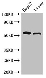
Figure 1. Western blot analysis of Scavenging Receptor SRB2/SCARB2 using anti-Scavenging Receptor SRB2/SCARB2 antibody (A05090-1). Electrophoresis was performed on a 5-20% SDS-PAGE gel at 70V (Stacking gel) / 90V (Resolving gel) for 2-3 hours. The sample well of each lane was loaded with 30 ug of sample under reducing conditions. Lane 1: human 293T whole cell lysates, Lane 2: human SH-SY5Y whole cell lysates. After electrophoresis, proteins were transferred to a nitrocellulose membrane at 150 mA for 50-90 minutes. Blocked the membrane with 5% non-fat milk/TBS for 1.5 hour at RT. The membrane was incubated with rabbit anti-Scavenging Receptor SRB2/SCARB2 antigen affinity purified polyclonal antibody (Catalog # A05090-1) at 0.5 microg/mL overnight at 4°C, then washed with TBS-0.1%Tween 3 times with 5 minutes each and probed with a goat anti-rabbit IgG-HRP secondary antibody at a dilution of 1:5000 for 1.5 hour at RT. The signal is developed using an Enhanced Chemiluminescent detection (ECL) kit (Catalog # EK1002) with Tanon 5200 system. A specific band was detected for Scavenging Receptor SRB2/SCARB2 at approximately 80 kDa. The expected band size for Scavenging Receptor SRB2/SCARB2 is at 54 kDa.
. Scavenging Receptor SRB2/SCARB2 was detected in a paraffin-embedded section of human lung cancer tissue. Heat mediated antigen retrieval was performed in EDTA buffer (pH 8.0, epitope retrieval solution). The tissue section was blocked with 10% goat serum. The tissue section was then incubated with 2 microg/ml rabbit anti-Scavenging Receptor SRB2/SCARB2 Antibody (A05090-1) overnight at 4°C. Peroxidase Conjugated Goat Anti-rabbit IgG was used as secondary antibody and incubated for 30 minutes at 37°C. The tissue section was developed using HRP Conjugated Rabbit IgG Super Vision Assay Kit (Catalog # SV0002) with DAB as the chromogen.")
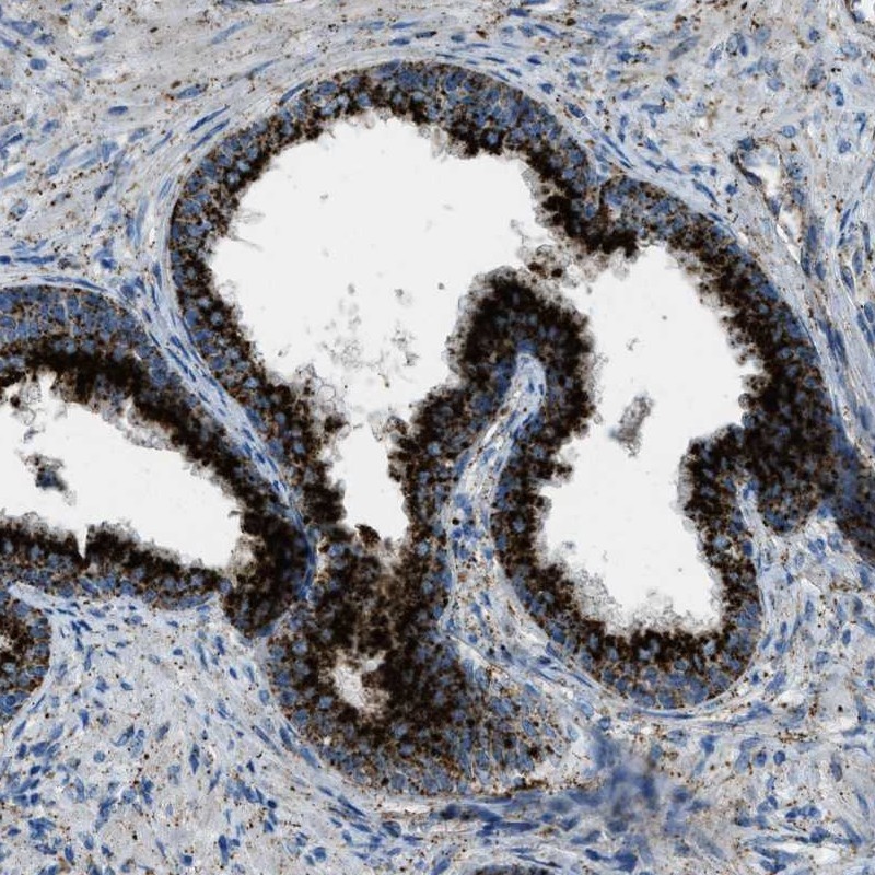
. Scavenging Receptor SRB2/SCARB2 was detected in a paraffin-embedded section of human prostate cancer tissue. Heat mediated antigen retrieval was performed in EDTA buffer (pH 8.0, epitope retrieval solution). The tissue section was blocked with 10% goat serum. The tissue section was then incubated with 2 microg/ml rabbit anti-Scavenging Receptor SRB2/SCARB2 Antibody (A05090-1) overnight at 4°C. Peroxidase Conjugated Goat Anti-rabbit IgG was used as secondary antibody and incubated for 30 minutes at 37°C. The tissue section was developed using HRP Conjugated Rabbit IgG Super Vision Assay Kit (Catalog # SV0002) with DAB as the chromogen.")
. Scavenging Receptor SRB2/SCARB2 was detected in a paraffin-embedded section of human endometrial cancer tissue. Heat mediated antigen retrieval was performed in EDTA buffer (pH 8.0, epitope retrieval solution). The tissue section was blocked with 10% goat serum. The tissue section was then incubated with 2 microg/ml rabbit anti-Scavenging Receptor SRB2/SCARB2 Antibody (A05090-1) overnight at 4°C. Peroxidase Conjugated Goat Anti-rabbit IgG was used as secondary antibody and incubated for 30 minutes at 37°C. The tissue section was developed using HRP Conjugated Rabbit IgG Super Vision Assay Kit (Catalog # SV0002) with DAB as the chromogen.")
. Scavenging Receptor SRB2/SCARB2 was detected in a paraffin-embedded section of human thyroid cancer tissue. Heat mediated antigen retrieval was performed in EDTA buffer (pH 8.0, epitope retrieval solution). The tissue section was blocked with 10% goat serum. The tissue section was then incubated with 2 microg/ml rabbit anti-Scavenging Receptor SRB2/SCARB2 Antibody (A05090-1) overnight at 4°C. Peroxidase Conjugated Goat Anti-rabbit IgG was used as secondary antibody and incubated for 30 minutes at 37°C. The tissue section was developed using HRP Conjugated Rabbit IgG Super Vision Assay Kit (Catalog # SV0002) with DAB as the chromogen.")
. Scavenging Receptor SRB2/SCARB2 was detected in a paraffin-embedded section of human breast cancer tissue. Heat mediated antigen retrieval was performed in EDTA buffer (pH 8.0, epitope retrieval solution). The tissue section was blocked with 10% goat serum. The tissue section was then incubated with 2 microg/ml rabbit anti-Scavenging Receptor SRB2/SCARB2 Antibody (A05090-1) overnight at 4°C. Peroxidase Conjugated Goat Anti-rabbit IgG was used as secondary antibody and incubated for 30 minutes at 37°C. The tissue section was developed using HRP Conjugated Rabbit IgG Super Vision Assay Kit (Catalog # SV0002) with DAB as the chromogen.")
. Scavenging Receptor SRB2/SCARB2 was detected in a paraffin-embedded section of human lung squamous cell carcinoma tissue. Heat mediated antigen retrieval was performed in EDTA buffer (pH 8.0, epitope retrieval solution). The tissue section was blocked with 10% goat serum. The tissue section was then incubated with 5 microg/mL rabbit anti-Scavenging Receptor SRB2/SCARB2 Antibody (A05090-1) overnight at 4°C. Cy3 Conjugated Goat Anti-Rabbit IgG (BA1032) was used as secondary antibody at 1:500 dilution and incubated for 30 minutes at 37°C. The section was counterstained with DAPI. Visualize using a fluorescence microscope and filter sets appropriate for the label used.")
. Overlay histogram showing U87 cells stained with A05090-1 (Blue line). To facilitate intracellular staining, cells were fixed with 4% paraformaldehyde and permeabilized with permeabilization buffer. The cells were blocked with 10% normal goat serum. And then incubated with rabbit anti-Scavenging Receptor SRB2/SCARB2 Antibody (A05090-1, 1 microg/1x106 cells) for 30 min at 20°C. DyLight®488 conjugated goat anti-rabbit IgG (BA1127, 5-10 microg/1x106 cells) was used as secondary antibody for 30 minutes at 20°C. Isotype control antibody (Green line) was rabbit IgG (1 microg/1x106) used under the same conditions. Unlabelled sample without incubation with primary antibody and secondary antibody (Red line) was used as a blank control.")
Figure 1. Western blot analysis of Scavenging Receptor SRB2/SCARB2 using anti-Scavenging Receptor SRB2/SCARB2 antibody (A05090-1). Electrophoresis was performed on a 5-20% SDS-PAGE gel at 70V (Stacking gel) / 90V (Resolving gel) for 2-3 hours. The sample well of each lane was loaded with 30 ug of sample under reducing conditions. Lane 1: human 293T whole cell lysates, Lane 2: human SH-SY5Y whole cell lysates. After electrophoresis, proteins were transferred to a nitrocellulose membrane at 150 mA for 50-90 minutes. Blocked the membrane with 5% non-fat milk/TBS for 1.5 hour at RT. The membrane was incubated with rabbit anti-Scavenging Receptor SRB2/SCARB2 antigen affinity purified polyclonal antibody (Catalog # A05090-1) at 0.5 microg/mL overnight at 4°C, then washed with TBS-0.1%Tween 3 times with 5 minutes each and probed with a goat anti-rabbit IgG-HRP secondary antibody at a dilution of 1:5000 for 1.5 hour at RT. The signal is developed using an Enhanced Chemiluminescent detection (ECL) kit (Catalog # EK1002) with Tanon 5200 system. A specific band was detected for Scavenging Receptor SRB2/SCARB2 at approximately 80 kDa. The expected band size for Scavenging Receptor SRB2/SCARB2 is at 54 kDa.
Anti-Scavenging Receptor SRB2/SCARB2 Antibody Picoband(r)
A05090-1-CARRIER-FREE
ApplicationsFlow Cytometry, ImmunoFluorescence, Western Blot, ELISA, ImmunoHistoChemistry
Product group Antibodies
ReactivityHuman
TargetSCARB2
Overview
- SupplierBoster Bio
- Product NameAnti-Scavenging Receptor SRB2/SCARB2 Antibody Picoband(r)
- Delivery Days Customer9
- ApplicationsFlow Cytometry, ImmunoFluorescence, Western Blot, ELISA, ImmunoHistoChemistry
- CertificationResearch Use Only
- ClonalityPolyclonal
- Concentration500 ug/ml
- Gene ID950
- Target nameSCARB2
- Target descriptionscavenger receptor class B member 2
- Target synonymsAMRF, CD36L2, EPM4, HLGP85, LGP85, LIMP-2, LIMPII, SR-BII, lysosome membrane protein 2, 85 kDa lysosomal membrane sialoglycoprotein, 85 kDa lysosomal sialoglycoprotein scavenger receptor class B, member 2, CD36 antigen (collagen type I receptor, thrombospondin receptor)-like 2 (lysosomal integral membrane protein II), CD36 antigen-like 2, LIMP II, lysosome membrane protein II
- HostRabbit
- IsotypeIgG
- Protein IDQ14108
- Protein NameLysosome membrane protein 2
- Scientific DescriptionBoster Bio Anti-Scavenging Receptor SRB2/SCARB2 Antibody Picoband® catalog # A05090-1. Tested in ELISA, Flow Cytometry, IF, IHC, WB applications. This antibody reacts with Human. The brand Picoband indicates this is a premium antibody that guarantees superior quality, high affinity, and strong signals with minimal background in Western blot applications. Only our best-performing antibodies are designated as Picoband, ensuring unmatched performance.
- ReactivityHuman
- Storage Instruction-20°C,2°C to 8°C
- UNSPSC12352203
Related products
Product group Antibodies
SCARB2 AntibodyCSB-PA619859LA01HU
ApplicationsImmunoFluorescence, Western Blot, ELISA, ImmunoHistoChemistry
ReactivityHuman, Mouse
TargetSCARB2
- SizePrice
Product group Antibodies

LIMP2 AntibodyABX430331
ApplicationsWestern Blot, ELISA, ImmunoHistoChemistry
- SizePrice

Product group Antibodies
Anti-SCARB2 AntibodyA31268
ApplicationsWestern Blot, ImmunoHistoChemistry
ReactivityHuman, Mouse, Rat
- SizePrice
Product group Antibodies

LIMPII / SCARB2 AntibodyLS-C747805
ApplicationsWestern Blot
ReactivityHuman, Mouse, Rat
TargetSCARB2
- SizePrice
Product group Antibodies
TargetSCARB2
- SizePrice
Product group Antibodies

ApplicationsWestern Blot, ELISA, ImmunoHistoChemistry
ReactivityHuman, Mouse
TargetSCARB2
- SizePrice
Product group Antibodies
Anti-SCARB2 AntibodyHPA018014
ApplicationsWestern Blot, ImmunoHistoChemistry
ReactivityHuman
TargetSCARB2
- SizePrice
Product group Antibodies
Scarb2 Polyclonal AntibodyCAC07916
ApplicationsImmunoFluorescence, Western Blot, ELISA, ImmunoHistoChemistry
ReactivityMouse
TargetSCARB2
- SizePrice