


Immunohistochemistry analysis in human kidney and lymph node tissues using Anti-SCIN antibody. Corresponding SCIN RNA-seq data are presented for the same tissues.

Immunohistochemistry analysis in human kidney and lymph node tissues using Anti-SCIN antibody. Corresponding SCIN RNA-seq data are presented for the same tissues.
Anti-SCIN Antibody
HPA020518
ApplicationsWestern Blot, ImmunoHistoChemistry
Product group Antibodies
ReactivityHuman
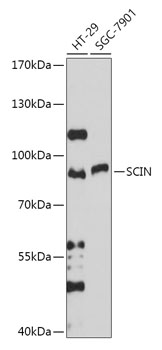
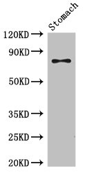
TargetSCIN
Overview
- SupplierAtlas Antibodies
- Product NameAnti-SCIN Antibody
- Delivery Days Customer4
- ApplicationsWestern Blot, ImmunoHistoChemistry
- CertificationResearch Use Only
- ClonalityPolyclonal
- ConjugateUnconjugated
- Gene ID85477
- Target nameSCIN
- Target descriptionscinderin
- Target synonymsscinderin, adseverin
- HostRabbit
- IsotypeIgG
- Protein IDQ9Y6U3
- Protein NameScinderin
- Scientific DescriptionRecombinant Protein Epitope Signature Tag (PrEST) antigen sequence
- ReactivityHuman
- Storage Instruction-20°C,2°C to 8°C
- UNSPSC41116161
Datasheet
MSDS
Related products
Product group Antibodies
Anti-SCIN AntibodyA306993
ApplicationsImmunoFluorescence, Western Blot, ImmunoCytoChemistry
ReactivityHuman, Mouse
- SizePrice

Product group Antibodies
Anti-SCIN Antibody Picoband(r)A07463-CARRIER-FREE
ApplicationsWestern Blot, ELISA
ReactivityHuman, Mouse, Rat
TargetSCIN
- SizePrice
Product group Antibodies
Anti-SCIN Antibody144-65171
ApplicationsImmunoFluorescence, Western Blot
ReactivityHuman, Mouse
TargetSCIN
- SizePrice
Product group Antibodies
Scin Polyclonal AntibodyCAC08144
ApplicationsImmunoFluorescence, Western Blot, ELISA
ReactivityMouse
TargetSCIN
- SizePrice
Product group Antibodies
SCIN AntibodyCSB-PA020823LA01HU
ApplicationsImmunoFluorescence, Western Blot, ELISA
ReactivityHuman, Mouse
TargetSCIN
- SizePrice
Product group Antibodies
SCIN Polyclonal AntibodyBS-17286R
ApplicationsImmunoFluorescence, Western Blot, ImmunoHistoChemistry, ImmunoHistoChemistry Frozen, ImmunoHistoChemistry Paraffin
ReactivityHuman, Mouse, Rat
TargetSCIN
- SizePrice
Product group Antibodies

SCIN Antibody (FITC)LS-C499726
ApplicationsELISA
ReactivityHuman
TargetSCIN
- SizePrice

Product group Antibodies
Anti-SCIN AntibodyHPA024264
ApplicationsImmunoHistoChemistry
ReactivityHuman
TargetSCIN
- SizePrice