


Figure 1. Western blot analysis of SCYL1 using anti-SCYL1 antibody (A04750-1). Electrophoresis was performed on a 5-20% SDS-PAGE gel at 70V (Stacking gel) / 90V (Resolving gel) for 2-3 hours. The sample well of each lane was loaded with 30 ug of sample under reducing conditions. Lane 1: human Hela whole cell lysates, Lane 2: human MCF-7 whole cell lysates, Lane 3: human T-47D whole cell lysates, Lane 4: human PC-3 whole cell lysates. After electrophoresis, proteins were transferred to a nitrocellulose membrane at 150 mA for 50-90 minutes. Blocked the membrane with 5% non-fat milk/TBS for 1.5 hour at RT. The membrane was incubated with rabbit anti-SCYL1 antigen affinity purified polyclonal antibody (Catalog # A04750-1) at 0.5 microg/mL overnight at 4°C, then washed with TBS-0.1%Tween 3 times with 5 minutes each and probed with a goat anti-rabbit IgG-HRP secondary antibody at a dilution of 1:5000 for 1.5 hour at RT. The signal is developed using an Enhanced Chemiluminescent detection (ECL) kit (Catalog # EK1002) with Tanon 5200 system. A specific band was detected for SCYL1 at approximately 115 kDa. The expected band size for SCYL1 is at 90 kDa.
. Overlay histogram showing HL-60 cells stained with A04750-1 (Blue line). To facilitate intracellular staining, cells were fixed with 4% paraformaldehyde and permeabilized with permeabilization buffer. The cells were blocked with 10% normal goat serum. And then incubated with rabbit anti-SCYL1 Antibody (A04750-1, 1 microg/1x106 cells) for 30 min at 20°C. DyLight®488 conjugated goat anti-rabbit IgG (BA1127, 5-10 microg/1x106 cells) was used as secondary antibody for 30 minutes at 20°C. Isotype control antibody (Green line) was rabbit IgG (1 microg/1x106) used under the same conditions. Unlabelled sample without incubation with primary antibody and secondary antibody (Red line) was used as a blank control.")
Figure 1. Western blot analysis of SCYL1 using anti-SCYL1 antibody (A04750-1). Electrophoresis was performed on a 5-20% SDS-PAGE gel at 70V (Stacking gel) / 90V (Resolving gel) for 2-3 hours. The sample well of each lane was loaded with 30 ug of sample under reducing conditions. Lane 1: human Hela whole cell lysates, Lane 2: human MCF-7 whole cell lysates, Lane 3: human T-47D whole cell lysates, Lane 4: human PC-3 whole cell lysates. After electrophoresis, proteins were transferred to a nitrocellulose membrane at 150 mA for 50-90 minutes. Blocked the membrane with 5% non-fat milk/TBS for 1.5 hour at RT. The membrane was incubated with rabbit anti-SCYL1 antigen affinity purified polyclonal antibody (Catalog # A04750-1) at 0.5 microg/mL overnight at 4°C, then washed with TBS-0.1%Tween 3 times with 5 minutes each and probed with a goat anti-rabbit IgG-HRP secondary antibody at a dilution of 1:5000 for 1.5 hour at RT. The signal is developed using an Enhanced Chemiluminescent detection (ECL) kit (Catalog # EK1002) with Tanon 5200 system. A specific band was detected for SCYL1 at approximately 115 kDa. The expected band size for SCYL1 is at 90 kDa.
Anti-SCYL1 Antibody Picoband(r)
A04750-1-CARRIER-FREE
ApplicationsFlow Cytometry, Western Blot, ELISA
Product group Antibodies
ReactivityHuman
TargetSCYL1
Overview
- SupplierBoster Bio
- Product NameAnti-SCYL1 Antibody Picoband(r)
- Delivery Days Customer9
- ApplicationsFlow Cytometry, Western Blot, ELISA
- CertificationResearch Use Only
- ClonalityPolyclonal
- Concentration500 ug/ml
- Gene ID57410
- Target nameSCYL1
- Target descriptionSCY1 like pseudokinase 1
- Target synonymsGKLP, HT019, NKTL, NTKL, P105, SCAR21, TAPK, TEIF, TRAP, N-terminal kinase-like protein, SCY1-like protein 1, SCY1-like, kinase-like 1, coated vesicle-associated kinase of 90 kDa, likely ortholog of mouse N-terminal kinase-like protein, telomerase regulation-associated protein, telomerase transcriptional elements-interacting factor, teratoma-associated tyrosine kinase
- HostRabbit
- IsotypeIgG
- Protein IDQ96KG9
- Protein NameN-terminal kinase-like protein
- Scientific DescriptionBoster Bio Anti-SCYL1 Antibody Picoband® catalog # A04750-1. Tested in ELISA, Flow Cytometry, WB applications. This antibody reacts with Human. The brand Picoband indicates this is a premium antibody that guarantees superior quality, high affinity, and strong signals with minimal background in Western blot applications. Only our best-performing antibodies are designated as Picoband, ensuring unmatched performance.
- ReactivityHuman
- Storage Instruction-20°C,2°C to 8°C
- UNSPSC12352203
Related products
Product group Antibodies
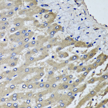
Anti-SCYL1 AntibodyA31561
ApplicationsImmunoFluorescence, Western Blot, ImmunoHistoChemistry
ReactivityHuman
- SizePrice
Product group Antibodies
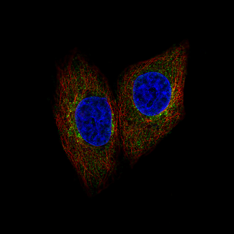
Anti-SCYL1 AntibodyHPA015015
ApplicationsImmunoCytoChemistry
ReactivityHuman
TargetSCYL1
- SizePrice
Product group Antibodies

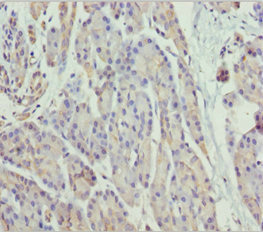
SCYL1 / NTKL AntibodyLS-C482692
ApplicationsImmunoFluorescence, Western Blot, ImmunoCytoChemistry, ImmunoHistoChemistry, ImmunoHistoChemistry Paraffin
ReactivityHuman
TargetSCYL1
- SizePrice
Product group Antibodies
SCYL1 AntibodyCSB-PA839356DSR1HU
ApplicationsELISA, ImmunoHistoChemistry
ReactivityHuman
TargetSCYL1
- SizePrice
Product group Antibodies
Scyl1 Polyclonal AntibodyCAC10793
ApplicationsELISA, ImmunoHistoChemistry
TargetSCYL1
- SizePrice
Product group Antibodies
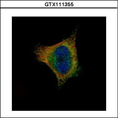
SCYL1 antibody [N3C2], InternalGTX111355
ApplicationsImmunoFluorescence, Western Blot, ImmunoCytoChemistry
ReactivityHuman
TargetSCYL1
- SizePrice
Product group Antibodies
Anti-SCYL1 Antibody144-06735
ApplicationsImmunoFluorescence, Western Blot, ImmunoHistoChemistry
ReactivityHuman, Mouse, Rat
TargetSCYL1
- SizePrice