

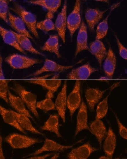
Anti-SEMA3C Antibody
144-61028
ApplicationsWestern Blot
Product group Antibodies
ReactivityHuman, Mouse, Rat
TargetSEMA3C
Overview
- SupplierRayBiotech
- Product NameAnti-SEMA3C Antibody
- Delivery Days Customer16
- ApplicationsWestern Blot
- CertificationResearch Use Only
- ClonalityPolyclonal
- ConjugateUnconjugated
- Gene ID10512
- Target nameSEMA3C
- Target descriptionsemaphorin 3C
- Target synonymsSEMAE, SemE, semaphorin-3C, sema E, sema domain, immunoglobulin domain (Ig), short basic domain, secreted, (semaphorin) 3C, semaphorin E
- HostRabbit
- IsotypeIgG
- Protein IDQ99985
- Protein NameSemaphorin-3C
- Scientific DescriptionSEMA3C Polyclonal Antibody
- ReactivityHuman, Mouse, Rat
- Storage Instruction-20°C
- UNSPSC12352203
Related products

Product group Antibodies
Anti-Semaphorin 3c/SEMA3C Antibody Picoband(r)A06444-1-CARRIER-FREE
ApplicationsWestern Blot, ELISA
ReactivityHuman, Mouse, Rat
TargetSEMA3C
- SizePrice
Product group Antibodies
Sema3C Polyclonal AntibodyCAC11467
ApplicationsELISA, ImmunoHistoChemistry
TargetSEMA3C
- SizePrice

Product group Antibodies
Sema3C Polyclonal AntibodyBS-6692R
ApplicationsImmunoFluorescence, Western Blot, ELISA, ImmunoCytoChemistry, ImmunoHistoChemistry, ImmunoHistoChemistry Frozen, ImmunoHistoChemistry Paraffin
ReactivityBovine, Chicken, Equine, Goat, Human, Mouse, Porcine, Rabbit, Rat, Sheep
TargetSEMA3C
- SizePrice
Product group Antibodies
SEMA3C AntibodyCSB-PA006602
ApplicationsELISA, ImmunoHistoChemistry
ReactivityHuman, Mouse, Rat
TargetSEMA3C
- SizePrice
Product group Antibodies
SEMA3C antibodyGTX66372
ApplicationsImmunoFluorescence, Western Blot, ImmunoCytoChemistry
ReactivityHuman, Mouse, Rat
TargetSEMA3C
- SizePrice
Product group Antibodies

ApplicationsWestern Blot
ReactivityHuman, Mouse, Rat
TargetSEMA3C
- SizePrice