


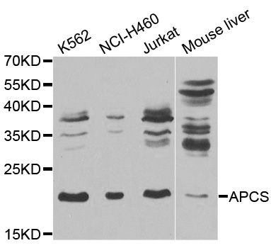
Figure 1. Western blot analysis of Serum Amyloid P/APCS using anti-Serum Amyloid P/APCS antibody (A00162). Electrophoresis was performed on a 5-20% SDS-PAGE gel at 70V (Stacking gel) / 90V (Resolving gel) for 2-3 hours. The sample well of each lane was loaded with 30 ug of sample under reducing conditions. Lane 1: human hepatocellular carcinoma tumor tissue (HCCT) lysates, Lane 2: human hepatocellular carcinoma paracancerous tissue (HCCP) lysates. After electrophoresis, proteins were transferred to a nitrocellulose membrane at 150 mA for 50-90 minutes. Blocked the membrane with 5% non-fat milk/TBS for 1.5 hour at RT. The membrane was incubated with rabbit anti-Serum Amyloid P/APCS antigen affinity purified polyclonal antibody (Catalog # A00162) at 0.5 microg/mL overnight at 4°C, then washed with TBS-0.1%Tween 3 times with 5 minutes each and probed with a goat anti-rabbit IgG-HRP secondary antibody at a dilution of 1:5000 for 1.5 hour at RT. The signal is developed using an Enhanced Chemiluminescent detection (ECL) kit (Catalog # EK1002) with Tanon 5200 system. A specific band was detected for Serum Amyloid P/APCS at approximately 25-29 kDa. The expected band size for Serum Amyloid P/APCS is at 25 kDa.
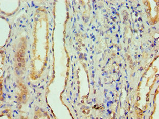
. Serum Amyloid P/APCS was detected in a paraffin-embedded section of human liver cancer tissue. Heat mediated antigen retrieval was performed in EDTA buffer (pH 8.0, epitope retrieval solution). The tissue section was blocked with 10% goat serum. The tissue section was then incubated with 2 microg/ml rabbit anti-Serum Amyloid P/APCS Antibody (A00162) overnight at 4°C. Peroxidase Conjugated Goat Anti-rabbit IgG was used as secondary antibody and incubated for 30 minutes at 37°C. The tissue section was developed using HRP Conjugated Rabbit IgG Super Vision Assay Kit (Catalog # SV0002) with DAB as the chromogen.")
. Serum Amyloid P/APCS was detected in a paraffin-embedded section of human liver cancer tissue. Heat mediated antigen retrieval was performed in EDTA buffer (pH 8.0, epitope retrieval solution). The tissue section was blocked with 10% goat serum. The tissue section was then incubated with 2 microg/ml rabbit anti-Serum Amyloid P/APCS Antibody (A00162) overnight at 4°C. Peroxidase Conjugated Goat Anti-rabbit IgG was used as secondary antibody and incubated for 30 minutes at 37°C. The tissue section was developed using HRP Conjugated Rabbit IgG Super Vision Assay Kit (Catalog # SV0002) with DAB as the chromogen.")
. Serum Amyloid P/APCS was detected in a paraffin-embedded section of human ovarian serous adenocarcinoma tissue. Heat mediated antigen retrieval was performed in EDTA buffer (pH 8.0, epitope retrieval solution). The tissue section was blocked with 10% goat serum. The tissue section was then incubated with 2 microg/ml rabbit anti-Serum Amyloid P/APCS Antibody (A00162) overnight at 4°C. Peroxidase Conjugated Goat Anti-rabbit IgG was used as secondary antibody and incubated for 30 minutes at 37°C. The tissue section was developed using HRP Conjugated Rabbit IgG Super Vision Assay Kit (Catalog # SV0002) with DAB as the chromogen.")
. Serum Amyloid P/APCS was detected in an immunocytochemical section of SiHa cells. Enzyme antigen retrieval was performed using IHC enzyme antigen retrieval reagent (AR0022) for 15 mins. The cells were blocked with 10% goat serum. And then incubated with 5 microg/mL rabbit anti-Serum Amyloid P/APCS Antibody (A00162) overnight at 4°C. DyLight488 Conjugated Goat Anti-Rabbit IgG (BA1127) was used as secondary antibody at 1:500 dilution and incubated for 30 minutes at 37°C. The section was counterstained with DAPI. Visualize using a fluorescence microscope and filter sets appropriate for the label used.")
. Overlay histogram showing HL-60 cells stained with A00162 (Blue line). To facilitate intracellular staining, cells were fixed with 4% paraformaldehyde and permeabilized with permeabilization buffer. The cells were blocked with 10% normal goat serum. And then incubated with rabbit anti-Serum Amyloid P/APCS Antibody (A00162, 1 microg/1x106 cells) for 30 min at 20°C. DyLight®488 conjugated goat anti-rabbit IgG (BA1127, 5-10 microg/1x106 cells) was used as secondary antibody for 30 minutes at 20°C. Isotype control antibody (Green line) was rabbit IgG (1 microg/1x106) used under the same conditions. Unlabelled sample without incubation with primary antibody and secondary antibody (Red line) was used as a blank control.")
Figure 1. Western blot analysis of Serum Amyloid P/APCS using anti-Serum Amyloid P/APCS antibody (A00162). Electrophoresis was performed on a 5-20% SDS-PAGE gel at 70V (Stacking gel) / 90V (Resolving gel) for 2-3 hours. The sample well of each lane was loaded with 30 ug of sample under reducing conditions. Lane 1: human hepatocellular carcinoma tumor tissue (HCCT) lysates, Lane 2: human hepatocellular carcinoma paracancerous tissue (HCCP) lysates. After electrophoresis, proteins were transferred to a nitrocellulose membrane at 150 mA for 50-90 minutes. Blocked the membrane with 5% non-fat milk/TBS for 1.5 hour at RT. The membrane was incubated with rabbit anti-Serum Amyloid P/APCS antigen affinity purified polyclonal antibody (Catalog # A00162) at 0.5 microg/mL overnight at 4°C, then washed with TBS-0.1%Tween 3 times with 5 minutes each and probed with a goat anti-rabbit IgG-HRP secondary antibody at a dilution of 1:5000 for 1.5 hour at RT. The signal is developed using an Enhanced Chemiluminescent detection (ECL) kit (Catalog # EK1002) with Tanon 5200 system. A specific band was detected for Serum Amyloid P/APCS at approximately 25-29 kDa. The expected band size for Serum Amyloid P/APCS is at 25 kDa.
Anti-Serum Amyloid P/APCS Antibody Picoband(r)
A00162-CARRIER-FREE
ApplicationsFlow Cytometry, ImmunoFluorescence, Western Blot, ELISA, ImmunoCytoChemistry, ImmunoHistoChemistry
Product group Antibodies
ReactivityHuman
TargetAPCS
Overview
- SupplierBoster Bio
- Product NameAnti-Serum Amyloid P/APCS Antibody Picoband(r)
- Delivery Days Customer9
- ApplicationsFlow Cytometry, ImmunoFluorescence, Western Blot, ELISA, ImmunoCytoChemistry, ImmunoHistoChemistry
- CertificationResearch Use Only
- ClonalityPolyclonal
- Concentration500 ug/ml
- Gene ID325
- Target nameAPCS
- Target descriptionamyloid P component, serum
- Target synonymsHEL-S-92n, PTX2, SAP, serum amyloid P-component, 9.5S alpha-1-glycoprotein, epididymis secretory sperm binding protein Li 92n, pentaxin-related, pentraxin-2, pentraxin-related
- HostRabbit
- IsotypeIgG
- Protein IDP02743
- Protein NameSerum amyloid P-component
- Scientific DescriptionBoster Bio Anti-Serum Amyloid P/APCS Antibody Picoband® catalog # A00162. Tested in ELISA, Flow Cytometry, IF, IHC, ICC, WB applications. This antibody reacts with Human. The brand Picoband indicates this is a premium antibody that guarantees superior quality, high affinity, and strong signals with minimal background in Western blot applications. Only our best-performing antibodies are designated as Picoband, ensuring unmatched performance.
- ReactivityHuman
- Storage Instruction-20°C,2°C to 8°C
- UNSPSC12352203
Related products
Product group Antibodies
Anti-APCS AntibodyA38484
ApplicationsWestern Blot, ImmunoHistoChemistry
ReactivityHuman
- SizePrice
Product group Antibodies
Anti-APCS Antibody144-65443
ApplicationsWestern Blot, ImmunoHistoChemistry
ReactivityHuman, Mouse, Rat
TargetAPCS
- SizePrice
Product group Antibodies
APCS Polyclonal AntibodyBS-3765R
ApplicationsImmunoFluorescence, Western Blot, ELISA, ImmunoCytoChemistry, ImmunoHistoChemistry, ImmunoHistoChemistry Frozen, ImmunoHistoChemistry Paraffin
ReactivityHuman, Mouse, Porcine, Rat
TargetAPCS
- SizePrice
Product group Antibodies
APCS AntibodyCSB-PA001898ESR1HU
ApplicationsELISA, ImmunoHistoChemistry
ReactivityHuman
TargetAPCS
- SizePrice
Product group Antibodies
ApplicationsImmunoPrecipitation, Western Blot, ImmunoCytoChemistry, ImmunoHistoChemistry
TargetAPCS
- SizePrice
Product group Antibodies
Serum Amyloid P antibodyGTX20688
ApplicationsImmunoElectrophoresis, ImmunoFluorescence, ELISA, ImmunoCytoChemistry, ImmunoHistoChemistry, Other Application
ReactivityHuman
TargetAPCS
- SizePrice

Product group Antibodies
Anti-APCS AntibodyHPA053294
ApplicationsWestern Blot, ImmunoHistoChemistry
ReactivityHuman
TargetAPCS
- SizePrice
Product group Antibodies

APCS / Serum Amyloid P / SAP AntibodyLS-C663198
ApplicationsWestern Blot
ReactivityMouse
TargetAPCS
- SizePrice