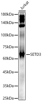
Bio-Connect
Anti-SETD3 Antibody

Anti-SETD3 Antibody

A307360
Antibodies.com
ApplicationsImmunoPrecipitation, Western Blot
Product group Antibodies
Sign in to order and to see your custom pricing.
Large volume orders?
Order with a bulk request

Overview

  • Supplier
    Antibodies.com
  • Product Name
    Anti-SETD3 Antibody
  • Delivery Days Customer
    7
  • Applications
    ImmunoPrecipitation, Western Blot
  • Certification
    Research Use Only
  • Clonality
    Polyclonal
  • Conjugate
    Unconjugated
  • Host
    Rabbit
  • Isotype
    IgG
  • Scientific Description
    Rabbit polyclonal antibody to SETD3.
  • UNSPSC
    12352203

Related products

Product group Antibodies
RayBiotech
ApplicationsImmunoPrecipitation, Western Blot
TargetSETD3
  • SizePrice
Product group Antibodies
Bethyl Laboratories
ApplicationsImmunoPrecipitation, Western Blot
TargetSETD3
  • SizePrice
Product group Antibodies
LifeSpan BioSciences
SETD3 AntibodyLS-C409610
ApplicationsWestern Blot
TargetSETD3
  • SizePrice
Product group Antibodies
Absolute Antibody
Anti-SETD3 [RAB-C384]AB01864-1.1-BT
ApplicationsImmunoFluorescence, ImmunoPrecipitation
TargetSETD3
  • SizePrice
Immunohistochemical staining of human small intestine shows strong cytoplasmic positivity in glandular cells.
Product group Antibodies
Atlas Antibodies
ApplicationsWestern Blot, ImmunoHistoChemistry
TargetSETD3
  • SizePrice
Figure 1. Western blot analysis of SETD3 using anti-SETD3 antibody (A10965-1). Electrophoresis was performed on a 5-20% SDS-PAGE gel at 70V (Stacking gel) / 90V (Resolving gel) for 2-3 hours. The sample well of each lane was loaded with 30 ug of sample under reducing conditions. Lane 1: human MCF-7 whole cell lysates, Lane 2: human T-47D whole cell lysates, Lane 3: human Jurkat whole cell lysates, Lane 4: human MOLT-4 whole cell lysates, Lane 5: human HL-60 whole cell lysates, Lane 6: human PC-3 whole cell lysates, Lane 7: human RT4 whole cell lysates. After electrophoresis, proteins were transferred to a nitrocellulose membrane at 150 mA for 50-90 minutes. Blocked the membrane with 5% non-fat milk/TBS for 1.5 hour at RT. The membrane was incubated with rabbit anti-SETD3 antigen affinity purified polyclonal antibody (Catalog # A10965-1) at 0.25 microg/mL overnight at 4°C, then washed with TBS-0.1%Tween 3 times with 5 minutes each and probed with a goat anti-rabbit IgG-HRP secondary antibody at a dilution of 1:5000 for 1.5 hour at RT. The signal is developed using an Enhanced Chemiluminescent detection (ECL) kit (Catalog # EK1002) with Tanon 5200 system. A specific band was detected for SETD3 at approximately 60 kDa. The expected band size for SETD3 is at 67 kDa.
Product group Antibodies
Boster Bio
Anti-SETD3 Antibody Picoband(r)A10965-1-CARRIER-FREE
ApplicationsFlow Cytometry, ImmunoFluorescence, Western Blot, ELISA, ImmunoCytoChemistry, ImmunoHistoChemistry
TargetSETD3
  • SizePrice
IHC image of CSB-PA769772LA01HU diluted at 1:400 and staining in paraffin-embedded human appendix tissue performed on a Leica BondTM system. After dewaxing and hydration, antigen retrieval was mediated by high pressure in a citrate buffer (pH 6.0). Section was blocked with 10% normal goat serum 30min at RT. Then primary antibody (1% BSA) was incubated at 4°C overnight. The primary is detected by a biotinylated secondary antibody and visualized using an HRP conjugated SP system.
Product group Antibodies
Cusabio
SETD3 AntibodyCSB-PA769772LA01HU
ApplicationsELISA, ImmunoHistoChemistry
ReactivityHuman
TargetSETD3
  • SizePrice