


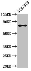
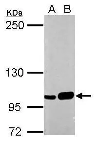
Figure 1. Western blot analysis of SFPQ using anti-SFPQ antibody (A02243-2). Electrophoresis was performed on a 5-20% SDS-PAGE gel at 70V (Stacking gel) / 90V (Resolving gel) for 2-3 hours. The sample well of each lane was loaded with 30 ug of sample under reducing conditions. Lane 1: human Hela whole cell lysates, Lane 2: human 293T whole cell lysates, Lane 3: human HepG2 whole cell lysates, Lane 4: human PC-3 whole cell lysates, Lane 5: rat brain tissue lysates, Lane 6: mouse brain tissue lysates, Lane 7: mouse C2C12 whole cell lysates, Lane 8: mouse NIH/3T3 whole cell lysates. After electrophoresis, proteins were transferred to a nitrocellulose membrane at 150 mA for 50-90 minutes. Blocked the membrane with 5% non-fat milk/TBS for 1.5 hour at RT. The membrane was incubated with rabbit anti-SFPQ antigen affinity purified polyclonal antibody (Catalog # A02243-2) at 0.5 microg/mL overnight at 4°C, then washed with TBS-0.1%Tween 3 times with 5 minutes each and probed with a goat anti-rabbit IgG-HRP secondary antibody at a dilution of 1:5000 for 1.5 hour at RT. The signal is developed using an Enhanced Chemiluminescent detection (ECL) kit (Catalog # EK1002) with Tanon 5200 system. A specific band was detected for SFPQ at approximately 100 kDa. The expected band size for SFPQ is at 76 kDa.
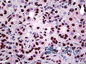
. SFPQ was detected in a paraffin-embedded section of human liver cancer tissue. Heat mediated antigen retrieval was performed in EDTA buffer (pH 8.0, epitope retrieval solution). The tissue section was blocked with 10% goat serum. The tissue section was then incubated with 2 microg/ml rabbit anti-SFPQ Antibody (A02243-2) overnight at 4°C. Peroxidase Conjugated Goat Anti-rabbit IgG was used as secondary antibody and incubated for 30 minutes at 37°C. The tissue section was developed using HRP Conjugated Rabbit IgG Super Vision Assay Kit (Catalog # SV0002) with DAB as the chromogen.")
. SFPQ was detected in a paraffin-embedded section of human ovarian cancer tissue. Heat mediated antigen retrieval was performed in EDTA buffer (pH 8.0, epitope retrieval solution). The tissue section was blocked with 10% goat serum. The tissue section was then incubated with 2 microg/ml rabbit anti-SFPQ Antibody (A02243-2) overnight at 4°C. Peroxidase Conjugated Goat Anti-rabbit IgG was used as secondary antibody and incubated for 30 minutes at 37°C. The tissue section was developed using HRP Conjugated Rabbit IgG Super Vision Assay Kit (Catalog # SV0002) with DAB as the chromogen.")
. SFPQ was detected in a paraffin-embedded section of human squamous cell carcinoma of the penis tissue. Heat mediated antigen retrieval was performed in EDTA buffer (pH 8.0, epitope retrieval solution). The tissue section was blocked with 10% goat serum. The tissue section was then incubated with 2 microg/ml rabbit anti-SFPQ Antibody (A02243-2) overnight at 4°C. Peroxidase Conjugated Goat Anti-rabbit IgG was used as secondary antibody and incubated for 30 minutes at 37°C. The tissue section was developed using HRP Conjugated Rabbit IgG Super Vision Assay Kit (Catalog # SV0002) with DAB as the chromogen.")
. SFPQ was detected in a paraffin-embedded section of mouse brain tissue. Heat mediated antigen retrieval was performed in EDTA buffer (pH 8.0, epitope retrieval solution). The tissue section was blocked with 10% goat serum. The tissue section was then incubated with 2 microg/ml rabbit anti-SFPQ Antibody (A02243-2) overnight at 4°C. Peroxidase Conjugated Goat Anti-rabbit IgG was used as secondary antibody and incubated for 30 minutes at 37°C. The tissue section was developed using HRP Conjugated Rabbit IgG Super Vision Assay Kit (Catalog # SV0002) with DAB as the chromogen.")
. SFPQ was detected in a paraffin-embedded section of rat brain tissue. Heat mediated antigen retrieval was performed in EDTA buffer (pH 8.0, epitope retrieval solution). The tissue section was blocked with 10% goat serum. The tissue section was then incubated with 2 microg/ml rabbit anti-SFPQ Antibody (A02243-2) overnight at 4°C. Peroxidase Conjugated Goat Anti-rabbit IgG was used as secondary antibody and incubated for 30 minutes at 37°C. The tissue section was developed using HRP Conjugated Rabbit IgG Super Vision Assay Kit (Catalog # SV0002) with DAB as the chromogen.")
 and anti-Tubulin Alpha antibody (M03989-3). SFPQ was detected in immunocytochemical section of MCF-7 cell. Enzyme antigen retrieval was performed using IHC enzyme antigen retrieval reagent (AR0022) for 15 mins. The cells were blocked with 10% goat serum. And then incubated with 5 microg/mL rabbit anti-SFPQ Antibody (A02243-2) and mouse anti-Tubulin Alpha antibody (M03989-3) overnight at 4°C. DyLight®488 Conjugated Goat Anti-Rabbit IgG (BA1127) and Cy3 Conjugated Goat Anti-Mouse IgG (BA1031) were used as secondary antibody at 1:100 dilution and incubated for 30 minutes at 37°C. The section was counterstained with DAPI. Visualize using a fluorescence microscope and filter sets appropriate for the label used.")
. Overlay histogram showing ANA-1 cells stained with A02243-2 (Blue line). To facilitate intracellular staining, cells were fixed with 4% paraformaldehyde and permeabilized with permeabilization buffer. The cells were blocked with 10% normal goat serum. And then incubated with rabbit anti-SFPQ Antibody (A02243-2, 1 microg/1x106 cells) for 30 min at 20°C. DyLight®488 conjugated goat anti-rabbit IgG (BA1127, 5-10 microg/1x106 cells) was used as secondary antibody for 30 minutes at 20°C. Isotype control antibody (Green line) was rabbit IgG (1 microg/1x106) used under the same conditions. Unlabelled sample without incubation with primary antibody and secondary antibody (Red line) was used as a blank control.")
. Overlay histogram showing RH35 cells stained with A02243-2 (Blue line). To facilitate intracellular staining, cells were fixed with 4% paraformaldehyde and permeabilized with permeabilization buffer. The cells were blocked with 10% normal goat serum. And then incubated with rabbit anti-SFPQ Antibody (A02243-2, 1 microg/1x106 cells) for 30 min at 20°C. DyLight®488 conjugated goat anti-rabbit IgG (BA1127, 5-10 microg/1x106 cells) was used as secondary antibody for 30 minutes at 20°C. Isotype control antibody (Green line) was rabbit IgG (1 microg/1x106) used under the same conditions. Unlabelled sample without incubation with primary antibody and secondary antibody (Red line) was used as a blank control.")
. Overlay histogram showing SiHa cells stained with A02243-2 (Blue line). To facilitate intracellular staining, cells were fixed with 4% paraformaldehyde and permeabilized with permeabilization buffer. The cells were blocked with 10% normal goat serum. And then incubated with rabbit anti-SFPQ Antibody (A02243-2, 1 microg/1x106 cells) for 30 min at 20°C. DyLight®488 conjugated goat anti-rabbit IgG (BA1127, 5-10 microg/1x106 cells) was used as secondary antibody for 30 minutes at 20°C. Isotype control antibody (Green line) was rabbit IgG (1 microg/1x106) used under the same conditions. Unlabelled sample without incubation with primary antibody and secondary antibody (Red line) was used as a blank control.")
Figure 1. Western blot analysis of SFPQ using anti-SFPQ antibody (A02243-2). Electrophoresis was performed on a 5-20% SDS-PAGE gel at 70V (Stacking gel) / 90V (Resolving gel) for 2-3 hours. The sample well of each lane was loaded with 30 ug of sample under reducing conditions. Lane 1: human Hela whole cell lysates, Lane 2: human 293T whole cell lysates, Lane 3: human HepG2 whole cell lysates, Lane 4: human PC-3 whole cell lysates, Lane 5: rat brain tissue lysates, Lane 6: mouse brain tissue lysates, Lane 7: mouse C2C12 whole cell lysates, Lane 8: mouse NIH/3T3 whole cell lysates. After electrophoresis, proteins were transferred to a nitrocellulose membrane at 150 mA for 50-90 minutes. Blocked the membrane with 5% non-fat milk/TBS for 1.5 hour at RT. The membrane was incubated with rabbit anti-SFPQ antigen affinity purified polyclonal antibody (Catalog # A02243-2) at 0.5 microg/mL overnight at 4°C, then washed with TBS-0.1%Tween 3 times with 5 minutes each and probed with a goat anti-rabbit IgG-HRP secondary antibody at a dilution of 1:5000 for 1.5 hour at RT. The signal is developed using an Enhanced Chemiluminescent detection (ECL) kit (Catalog # EK1002) with Tanon 5200 system. A specific band was detected for SFPQ at approximately 100 kDa. The expected band size for SFPQ is at 76 kDa.
Anti-SFPQ Antibody Picoband(r)
A02243-2-CARRIER-FREE
ApplicationsFlow Cytometry, ImmunoFluorescence, Western Blot, ImmunoCytoChemistry, ImmunoHistoChemistry
Product group Antibodies
ReactivityHuman, Mouse, Rat
TargetSFPQ
Overview
- SupplierBoster Bio
- Product NameAnti-SFPQ Antibody Picoband(r)
- Delivery Days Customer9
- ApplicationsFlow Cytometry, ImmunoFluorescence, Western Blot, ImmunoCytoChemistry, ImmunoHistoChemistry
- CertificationResearch Use Only
- ClonalityPolyclonal
- Concentration500 ug/ml
- Gene ID6421
- Target nameSFPQ
- Target descriptionsplicing factor proline and glutamine rich
- Target synonymsPOMP100, PPP1R140, PSF, splicing factor, proline- and glutamine-rich, 100 kDa DNA-pairing protein, DNA-binding p52/p100 complex, 100 kDa subunit, PTB-associated splicing factor, epididymis secretory sperm binding protein, polypyrimidine tract binding protein associated, polypyrimidine tract-binding protein-associated splicing factor, protein phosphatase 1, regulatory subunit 140, splicing factor proline/glutamine rich (polypyrimidine tract binding protein associated), splicing factor proline/glutamine-rich
- HostRabbit
- IsotypeIgG
- Protein IDP23246
- Protein NameSplicing factor, proline- and glutamine-rich
- Scientific DescriptionBoster Bio Anti-SFPQ Antibody Picoband® catalog # A02243-2. Tested in Flow Cytometry, IF, IHC, ICC, WB applications. This antibody reacts with Human, Mouse, Rat. The brand Picoband indicates this is a premium antibody that guarantees superior quality, high affinity, and strong signals with minimal background in Western blot applications. Only our best-performing antibodies are designated as Picoband, ensuring unmatched performance.
- ReactivityHuman, Mouse, Rat
- Storage Instruction-20°C,2°C to 8°C
- UNSPSC12352203
Related products
Product group Antibodies
SFPQ AntibodyCSB-PA021136LA01HU
ApplicationsWestern Blot, ELISA
ReactivityHuman, Mouse
TargetSFPQ
- SizePrice
Product group Antibodies
Anti-SFPQ AntibodyA326292
ApplicationsELISA, ImmunoHistoChemistry
ReactivityHuman
- SizePrice
Product group Antibodies

Goat anti-PSF / SFPQEB09523
ApplicationsELISA, ImmunoHistoChemistry
ReactivityBovine, Canine, Human, Mouse, Rat
TargetSFPQ
- SizePrice

Product group Antibodies
Anti-SFPQ AntibodyHPA047513
ApplicationsWestern Blot, ImmunoCytoChemistry, ImmunoHistoChemistry
ReactivityHuman
TargetSFPQ
- SizePrice
Product group Antibodies

SFPQ AntibodyLS-C667992
ApplicationsWestern Blot
ReactivityHuman
TargetSFPQ
- SizePrice
Product group Antibodies
SFPQ Recombinant Antibody, AbBy Fluor-594 ConjugatedBSM-61746R-BF594
ApplicationsImmunoFluorescence, Western Blot
ReactivityHuman, Mouse, Rat
TargetSFPQ
- SizePrice
Product group Antibodies
SFPQ Polyclonal AntibodyCAC14992
ApplicationsWestern Blot, ELISA
ReactivityMouse
TargetSFPQ
- SizePrice
Product group Antibodies
SFPQ antibodyGTX112116
ApplicationsWestern Blot, ImmunoHistoChemistry, ImmunoHistoChemistry Paraffin
ReactivityHuman
TargetSFPQ
- SizePrice