


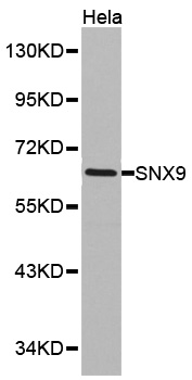
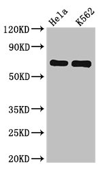
Figure 1. Western blot analysis of SH3PX1/SNX9 using anti-SH3PX1/SNX9 antibody (A03796-2). Electrophoresis was performed on a 5-20% SDS-PAGE gel at 70V (Stacking gel) / 90V (Resolving gel) for 2-3 hours. The sample well of each lane was loaded with 30 ug of sample under reducing conditions. Lane 1: human K562 whole cell lysates, Lane 2: human 293T whole cell lysates, Lane 3: human placenta tissue lysates. After electrophoresis, proteins were transferred to a nitrocellulose membrane at 150 mA for 50-90 minutes. Blocked the membrane with 5% non-fat milk/TBS for 1.5 hour at RT. The membrane was incubated with rabbit anti-SH3PX1/SNX9 antigen affinity purified polyclonal antibody (Catalog # A03796-2) at 0.25 microg/mL overnight at 4°C, then washed with TBS-0.1%Tween 3 times with 5 minutes each and probed with a goat anti-rabbit IgG-HRP secondary antibody at a dilution of 1:5000 for 1.5 hour at RT. The signal is developed using an Enhanced Chemiluminescent detection (ECL) kit (Catalog # EK1002) with Tanon 5200 system. A specific band was detected for SH3PX1/SNX9 at approximately 78 kDa. The expected band size for SH3PX1/SNX9 is at 67 kDa.
. Overlay histogram showing MCF-7 cells stained with A03796-2 (Blue line). To facilitate intracellular staining, cells were fixed with 4% paraformaldehyde and permeabilized with permeabilization buffer. The cells were blocked with 10% normal goat serum. And then incubated with rabbit anti-SH3PX1/SNX9 Antibody (A03796-2, 1 microg/1x106 cells) for 30 min at 20°C. DyLight®488 conjugated goat anti-rabbit IgG (BA1127, 5-10 microg/1x106 cells) was used as secondary antibody for 30 minutes at 20°C. Isotype control antibody (Green line) was rabbit IgG (1 microg/1x106) used under the same conditions. Unlabelled sample without incubation with primary antibody and secondary antibody (Red line) was used as a blank control.")
Figure 1. Western blot analysis of SH3PX1/SNX9 using anti-SH3PX1/SNX9 antibody (A03796-2). Electrophoresis was performed on a 5-20% SDS-PAGE gel at 70V (Stacking gel) / 90V (Resolving gel) for 2-3 hours. The sample well of each lane was loaded with 30 ug of sample under reducing conditions. Lane 1: human K562 whole cell lysates, Lane 2: human 293T whole cell lysates, Lane 3: human placenta tissue lysates. After electrophoresis, proteins were transferred to a nitrocellulose membrane at 150 mA for 50-90 minutes. Blocked the membrane with 5% non-fat milk/TBS for 1.5 hour at RT. The membrane was incubated with rabbit anti-SH3PX1/SNX9 antigen affinity purified polyclonal antibody (Catalog # A03796-2) at 0.25 microg/mL overnight at 4°C, then washed with TBS-0.1%Tween 3 times with 5 minutes each and probed with a goat anti-rabbit IgG-HRP secondary antibody at a dilution of 1:5000 for 1.5 hour at RT. The signal is developed using an Enhanced Chemiluminescent detection (ECL) kit (Catalog # EK1002) with Tanon 5200 system. A specific band was detected for SH3PX1/SNX9 at approximately 78 kDa. The expected band size for SH3PX1/SNX9 is at 67 kDa.
Anti-SH3PX1/SNX9 Antibody Picoband(r)
A03796-2-CARRIER-FREE
ApplicationsFlow Cytometry, Western Blot, ELISA
Product group Antibodies
ReactivityHuman
TargetSNX9
Overview
- SupplierBoster Bio
- Product NameAnti-SH3PX1/SNX9 Antibody Picoband(r)
- Delivery Days Customer9
- ApplicationsFlow Cytometry, Western Blot, ELISA
- CertificationResearch Use Only
- ClonalityPolyclonal
- Concentration500 ug/ml
- Gene ID51429
- Target nameSNX9
- Target descriptionsorting nexin 9
- Target synonymsSDP1, SH3PX1, SH3PXD3A, WISP, sorting nexin-9, SH3 and PX domain-containing protein 1, SH3 and PX domain-containing protein 3A, Wiskott-Aldrich syndrome protein (WASP) interactor protein
- HostRabbit
- IsotypeIgG
- Protein IDQ9Y5X1
- Protein NameSorting nexin-9
- Scientific DescriptionBoster Bio Anti-SH3PX1/SNX9 Antibody Picoband® catalog # A03796-2. Tested in ELISA, Flow Cytometry, WB applications. This antibody reacts with Human. The brand Picoband indicates this is a premium antibody that guarantees superior quality, high affinity, and strong signals with minimal background in Western blot applications. Only our best-performing antibodies are designated as Picoband, ensuring unmatched performance.
- ReactivityHuman
- Storage Instruction-20°C,2°C to 8°C
- UNSPSC12352203
Related products
Product group Antibodies
Anti-SNX9 AntibodyA43977
ApplicationsWestern Blot
ReactivityHuman, Mouse, Rat
- SizePrice
Product group Antibodies

Goat anti-SNX9EB07157
ApplicationsImmunoFluorescence, ELISA
ReactivityCanine, Human, Mouse
TargetSNX9
- SizePrice

Product group Antibodies
Anti-SNX9 AntibodyHPA031410
ApplicationsWestern Blot, ImmunoCytoChemistry, ImmunoHistoChemistry
ReactivityHuman, Mouse, Rat
TargetSNX9
- SizePrice
Product group Antibodies
SNX9 AntibodyCSB-PA896917LA01HU
ApplicationsImmunoFluorescence, Western Blot, ELISA, ImmunoHistoChemistry
ReactivityHuman
TargetSNX9
- SizePrice
Product group Antibodies

SNX9 / WISP AntibodyLS-C335116
ApplicationsWestern Blot, ImmunoHistoChemistry
ReactivityHuman, Mouse, Rat
TargetSNX9
- SizePrice
Product group Antibodies
Snx9 Polyclonal AntibodyCAC07836
ApplicationsImmunoFluorescence, Western Blot, ELISA, ImmunoHistoChemistry
TargetSNX9
- SizePrice
Product group Antibodies
SNX9 antibodyGTX103383
ApplicationsWestern Blot, ImmunoHistoChemistry, ImmunoHistoChemistry Paraffin
ReactivityHuman
TargetSNX9
- SizePrice
Product group Antibodies
SH3PX1 Polyclonal AntibodyBS-12407R
ApplicationsWestern Blot, ELISA
ReactivityBovine, Canine, Equine, Human, Mouse, Porcine, Rat
TargetSNX9
- SizePrice