


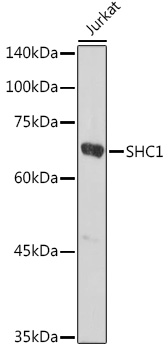
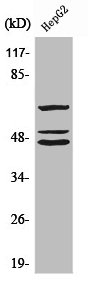
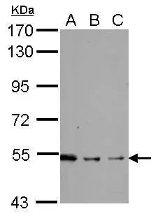
Figure 1. Western blot analysis of SHC using anti-SHC antibody (PB9391). Electrophoresis was performed on a 5-20% SDS-PAGE gel at 70V (Stacking gel) / 90V (Resolving gel) for 2-3 hours. The sample well of each lane was loaded with 50ug of sample under reducing conditions. Lane 1: human A431 whole cell lysate, Lane 2: human Hela whole cell lysate, Lane 3: human HepG2 whole cell lysate, Lane 4: human Jurkat whole cell lysate, Lane 5: human K562 whole cell lysate, Lane 6: human THP-1 whole cell lysate, Lane 7: rat C6 whole cell lysate, Lane 8: mouse thymus tissue lysate, Lane 9: mouse RAW246.7 whole cell lysate, Lane 10: mouse NIH3T3 whole cell lysate. After Electrophoresis, proteins were transferred to a Nitrocellulose membrane at 150mA for 50-90 minutes. Blocked the membrane with 5% Non-fat Milk/ TBS for 1.5 hour at RT. The membrane was incubated with rabbit anti-SHC antigen affinity purified polyclonal antibody (Catalog # PB9391) at 0.5 microg/mL overnight at 4°C, then washed with TBS-0.1%Tween 3 times with 5 minutes each and probed with a goat anti-rabbit IgG-HRP secondary antibody at a dilution of 1:10000 for 1.5 hour at RT. The signal is developed using an Enhanced Chemiluminescent detection (ECL) kit (Catalog # EK1002) with Tanon 5200 system. Specific bands were detected for SHC at approximately 46, 52, 66KD. The expected band size for SHC are at 46, 52, 66KD.
. SHC1 was detected in paraffin-embedded section of Mouse Brain Tissue. Heat mediated antigen retrieval was performed in citrate buffer (pH6, epitope retrieval solution) for 20 mins. The tissue section was blocked with 10% goat serum. The tissue section was then incubated with 1microg/ml rabbit anti-SHC1 Antibody (PB9391) overnight at 4°C. Biotinylated goat anti-rabbit IgG was used as secondary antibody and incubated for 30 minutes at 37°C. The tissue section was developed using Strepavidin-Biotin-Complex (SABC)(Catalog # SA1022) with DAB as the chromogen.")
. SHC1 was detected in paraffin-embedded section of Rat Brain Tissue. Heat mediated antigen retrieval was performed in citrate buffer (pH6, epitope retrieval solution) for 20 mins. The tissue section was blocked with 10% goat serum. The tissue section was then incubated with 1microg/ml rabbit anti-SHC1 Antibody (PB9391) overnight at 4°C. Biotinylated goat anti-rabbit IgG was used as secondary antibody and incubated for 30 minutes at 37°C. The tissue section was developed using Strepavidin-Biotin-Complex (SABC)(Catalog # SA1022) with DAB as the chromogen.")
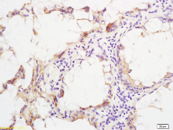
. SHC1 was detected in paraffin-embedded section of Human Lung Cancer Tissue. Heat mediated antigen retrieval was performed in citrate buffer (pH6, epitope retrieval solution) for 20 mins. The tissue section was blocked with 10% goat serum. The tissue section was then incubated with 1microg/ml rabbit anti-SHC1 Antibody (PB9391) overnight at 4°C. Biotinylated goat anti-rabbit IgG was used as secondary antibody and incubated for 30 minutes at 37°C. The tissue section was developed using Strepavidin-Biotin-Complex (SABC)(Catalog # SA1022) with DAB as the chromogen.")
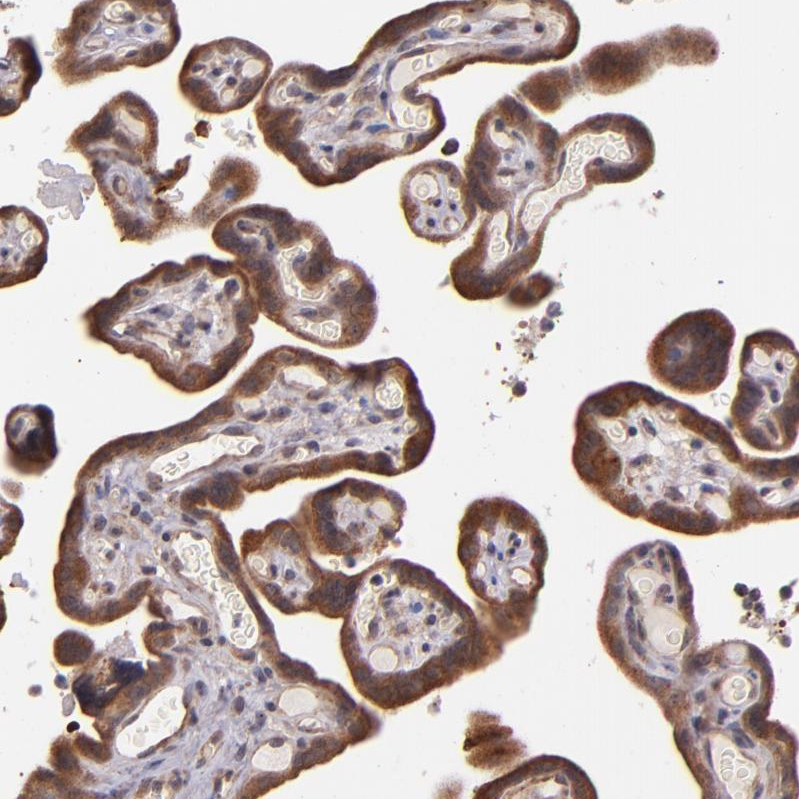
. SHC1 was detected in frozen section of Human Placenta Tissue. The tissue section was blocked with 10% goat serum. The tissue section was then incubated with 1microg/ml rabbit anti-SHC1 Antibody (PB9391) overnight at 4°C. Biotinylated goat anti-rabbit IgG was used as secondary antibody and incubated for 30 minutes at 37°C. The tissue section was developed using Strepavidin-Biotin-Complex (SABC)(Catalog # SA1022) with DAB as the chromogen.")
Figure 1. Western blot analysis of SHC using anti-SHC antibody (PB9391). Electrophoresis was performed on a 5-20% SDS-PAGE gel at 70V (Stacking gel) / 90V (Resolving gel) for 2-3 hours. The sample well of each lane was loaded with 50ug of sample under reducing conditions. Lane 1: human A431 whole cell lysate, Lane 2: human Hela whole cell lysate, Lane 3: human HepG2 whole cell lysate, Lane 4: human Jurkat whole cell lysate, Lane 5: human K562 whole cell lysate, Lane 6: human THP-1 whole cell lysate, Lane 7: rat C6 whole cell lysate, Lane 8: mouse thymus tissue lysate, Lane 9: mouse RAW246.7 whole cell lysate, Lane 10: mouse NIH3T3 whole cell lysate. After Electrophoresis, proteins were transferred to a Nitrocellulose membrane at 150mA for 50-90 minutes. Blocked the membrane with 5% Non-fat Milk/ TBS for 1.5 hour at RT. The membrane was incubated with rabbit anti-SHC antigen affinity purified polyclonal antibody (Catalog # PB9391) at 0.5 microg/mL overnight at 4°C, then washed with TBS-0.1%Tween 3 times with 5 minutes each and probed with a goat anti-rabbit IgG-HRP secondary antibody at a dilution of 1:10000 for 1.5 hour at RT. The signal is developed using an Enhanced Chemiluminescent detection (ECL) kit (Catalog # EK1002) with Tanon 5200 system. Specific bands were detected for SHC at approximately 46, 52, 66KD. The expected band size for SHC are at 46, 52, 66KD.
Anti-SHC/SHC1 Antibody Picoband(r)
PB9391-CARRIER-FREE
ApplicationsWestern Blot, ImmunoHistoChemistry, ImmunoHistoChemistry Frozen
Product group Antibodies
ReactivityHamster, Human, Mouse, Rat
TargetSHC1
Overview
- SupplierBoster Bio
- Product NameAnti-SHC/SHC1 Antibody Picoband(r)
- Delivery Days Customer9
- Application Supplier NoteWB: The detection limit for SHC is approximately 0.1ng/lane under reducing conditions. Tested Species: In-house tested species with positive results. By Heat: Boiling the paraffin sections in 10mM citrate buffer, pH6.0, for 20mins is required for the staining of formalin/paraffin sections. Other applications have not been tested. Optimal dilutions should be determined by end users.
- ApplicationsWestern Blot, ImmunoHistoChemistry, ImmunoHistoChemistry Frozen
- CertificationResearch Use Only
- ClonalityPolyclonal
- Concentration500 ug/ml
- Gene ID6464
- Target nameSHC1
- Target descriptionSHC adaptor protein 1
- Target synonymsSHC, SHCA, SHC-transforming protein 1, SH2 domain protein C1, SHC (Src homology 2 domain containing) transforming protein 1, SHC-transforming protein 3, SHC-transforming protein A
- HostRabbit
- IsotypeIgG
- Protein IDP29353
- Protein NameSHC-transforming protein 1
- Scientific DescriptionBoster Bio Anti-SHC/SHC1 Antibody Picoband® catalog # PB9391. Tested in IHC, IHC-F, WB applications. This antibody reacts with Human, Mouse, Rat. The brand Picoband indicates this is a premium antibody that guarantees superior quality, high affinity, and strong signals with minimal background in Western blot applications. Only our best-performing antibodies are designated as Picoband, ensuring unmatched performance.
- ReactivityHamster, Human, Mouse, Rat
- Storage Instruction-20°C,2°C to 8°C
- UNSPSC12352203
Related products
Product group Antibodies
Anti-SHC AntibodyA16912
ApplicationsImmunoFluorescence, Western Blot, ImmunoCytoChemistry, ImmunoHistoChemistry
ReactivityHuman, Mouse, Rat
- SizePrice
Product group Antibodies
Anti-SHC1 Antibody144-07725
ApplicationsImmunoFluorescence, Western Blot, ImmunoHistoChemistry
ReactivityHuman, Mouse, Rat
TargetSHC1
- SizePrice
Product group Antibodies
References
ApplicationsImmunoFluorescence, Western Blot, ELISA, ImmunoHistoChemistry, ImmunoHistoChemistry Frozen, ImmunoHistoChemistry Paraffin
ReactivityHuman, Mouse, Rat
TargetSHC1
- SizePrice
Product group Antibodies
SHC1 AntibodyCSB-PA004082
ApplicationsWestern Blot, ELISA, ImmunoHistoChemistry
ReactivityHuman, Mouse, Rat
TargetSHC1
- SizePrice
Product group Antibodies
ApplicationsImmunoPrecipitation, Western Blot, ImmunoCytoChemistry, ImmunoHistoChemistry
TargetSHC1
- SizePrice
Product group Antibodies

p66 / SHC AntibodyLS-C409277
ApplicationsWestern Blot, ImmunoHistoChemistry
ReactivityHuman, Mouse, Rat
TargetSHC1
- SizePrice
Product group Antibodies
Anti-SHC1 AntibodyHPA001844
ApplicationsImmunoHistoChemistry
ReactivityHuman
TargetSHC1
- SizePrice
Product group Antibodies
SHC1 antibody [N3C2], InternalGTX113267
ApplicationsWestern Blot
ReactivityHuman, Mouse
TargetSHC1
- SizePrice
Product group Antibodies
Anti-SHC1 AntibodyCAB7725
ApplicationsImmunoFluorescence, Western Blot, ELISA, ImmunoCytoChemistry, ImmunoHistoChemistry, ImmunoHistoChemistry Paraffin
ReactivityHuman
TargetSHC1
- SizePrice