


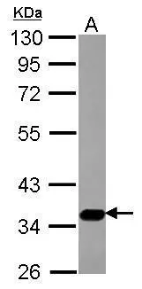
Figure 1. Western blot analysis of SLC17A5 using anti-SLC17A5 antibody (A06657-2). Electrophoresis was performed on a 5-20% SDS-PAGE gel at 70V (Stacking gel) / 90V (Resolving gel) for 2-3 hours. The sample well of each lane was loaded with 30 ug of sample unde r reducing conditions. Lane 1: human Jurkat whole cell lysates, Lane 2: human 293T whole cell lysates, Lane 3: monkey COS-7 whole cell lysates, Lane 4: human THP-1 whole cell lysates, Lane 5: rat NRK whole cell lysates, Lane 6: mouse HBZY-1 whole cell lysates. After electrophoresis, proteins were transferred to a nitrocellulose membrane at 150 mA for 50-90 minutes. Blocked the membrane with 5% non-fat milk/TBS for 1.5 hour at RT. The membrane was incubated with rabbit anti-SLC17A5 antigen affinity purified polyclonal antibody (Catalog # A06657-2) at 0.5 microg/mL overnight at 4°C, then washed with TBS-0.1%Tween 3 times with 5 minutes each and probed with a goat anti-rabbit IgG-HRP secondary antibody at a dilution of 1:5000 for 1.5 hour at RT. The signal is developed using an Enhanced Chemiluminescent detection (ECL) kit (Catalog # EK1002) with Tanon 5200 system. A specific band was detected for SLC17A5 at approximately 36 kDa. The expected band size for SLC17A5 is at 55 kDa.
. SLC17A5 was detected in a paraffin-embedded section of human thyroid cancer tissue. Heat mediated antigen retrieval was performed in EDTA buffer (pH 8.0, epitope retrieval solution). The tissue section was blocked with 10% goat serum. The tissue section was then incubated with 2 microg/ml rabbit anti-SLC17A5 Antibody (A06657-2) overnight at 4°C. Peroxidase Conjugated Goat Anti-rabbit IgG was used as secondary antibody and incubated for 30 minutes at 37°C. The tissue section was developed using HRP Conjugated Rabbit IgG Super Vision Assay Kit (Catalog # SV0002) with DAB as the chromogen.")
Figure 1. Western blot analysis of SLC17A5 using anti-SLC17A5 antibody (A06657-2). Electrophoresis was performed on a 5-20% SDS-PAGE gel at 70V (Stacking gel) / 90V (Resolving gel) for 2-3 hours. The sample well of each lane was loaded with 30 ug of sample unde r reducing conditions. Lane 1: human Jurkat whole cell lysates, Lane 2: human 293T whole cell lysates, Lane 3: monkey COS-7 whole cell lysates, Lane 4: human THP-1 whole cell lysates, Lane 5: rat NRK whole cell lysates, Lane 6: mouse HBZY-1 whole cell lysates. After electrophoresis, proteins were transferred to a nitrocellulose membrane at 150 mA for 50-90 minutes. Blocked the membrane with 5% non-fat milk/TBS for 1.5 hour at RT. The membrane was incubated with rabbit anti-SLC17A5 antigen affinity purified polyclonal antibody (Catalog # A06657-2) at 0.5 microg/mL overnight at 4°C, then washed with TBS-0.1%Tween 3 times with 5 minutes each and probed with a goat anti-rabbit IgG-HRP secondary antibody at a dilution of 1:5000 for 1.5 hour at RT. The signal is developed using an Enhanced Chemiluminescent detection (ECL) kit (Catalog # EK1002) with Tanon 5200 system. A specific band was detected for SLC17A5 at approximately 36 kDa. The expected band size for SLC17A5 is at 55 kDa.
Anti-SLC17A5 Antibody Picoband(r)
A06657-2-CARRIER-FREE
ApplicationsWestern Blot, ELISA, ImmunoHistoChemistry
Product group Antibodies
ReactivityHuman, Monkey, Mouse, Rat
TargetSLC17A5
Overview
- SupplierBoster Bio
- Product NameAnti-SLC17A5 Antibody Picoband(r)
- Delivery Days Customer9
- ApplicationsWestern Blot, ELISA, ImmunoHistoChemistry
- CertificationResearch Use Only
- ClonalityPolyclonal
- Concentration500 ug/ml
- Gene ID26503
- Target nameSLC17A5
- Target descriptionsolute carrier family 17 member 5
- Target synonymsAST, ISSD, NSD, SD, SIALIN, SIASD, SLD, VEAT, sialin, H(+)/nitrate cotransporter, H(+)/sialic acid cotransporter, acidic sugar transporter, infantile sialic acid storage disorder, membrane glycoprotein HP59, sialic acid storage disease, sodium/sialic acid cotransporter, solute carrier family 17 (acidic sugar transporter), member 5, solute carrier family 17 (anion/sugar transporter), member 5, vesicular H(+)/Aspartate-glutamate cotransporter, vesicular excitatory amino acid transporter
- HostRabbit
- IsotypeIgG
- Protein IDQ9NRA2
- Protein NameSialin
- Scientific DescriptionBoster Bio Anti-SLC17A5 Antibody Picoband® catalog # A06657-2. Tested in ELISA, IHC, WB applications. This antibody reacts with Human, Monkey, Mouse, Rat. The brand Picoband indicates this is a premium antibody that guarantees superior quality, high affinity, and strong signals with minimal background in Western blot applications. Only our best-performing antibodies are designated as Picoband, ensuring unmatched performance.
- ReactivityHuman, Monkey, Mouse, Rat
- Storage Instruction-20°C,2°C to 8°C
- UNSPSC12352203
Related products
Product group Antibodies
Anti-SLC17A5 Antibody144-63745
ApplicationsWestern Blot
ReactivityHuman, Mouse, Rat
TargetSLC17A5
- SizePrice
Product group Antibodies
References
SLC17A5 antibodyGTX115822
ApplicationsImmunoFluorescence, Western Blot, ImmunoCytoChemistry, ImmunoHistoChemistry, ImmunoHistoChemistry Paraffin
ReactivityHuman, Mouse
TargetSLC17A5
- SizePrice

Product group Antibodies
Anti-SLC17A5 AntibodyA89208
ApplicationsWestern Blot
ReactivityHuman, Rat
- SizePrice

Product group Antibodies

ApplicationsWestern Blot, ELISA
ReactivityHuman
TargetSLC17A5
- SizePrice
Product group Antibodies

SLC17A5 AntibodyLS-C750394
ApplicationsWestern Blot
ReactivityHuman, Rat
TargetSLC17A5
- SizePrice
Product group Antibodies
SLC17A5 AntibodyCSB-PA021421GA01HU
ApplicationsWestern Blot, ELISA
ReactivityHuman, Mouse, Rat
TargetSLC17A5
- SizePrice