


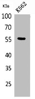
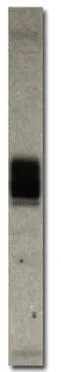
Figure 1. Western blot analysis of SLC2A5 using anti-SLC2A5 antibody (PB9960). Electrophoresis was performed on a 5-20% SDS-PAGE gel at 70V (Stacking gel) / 90V (Resolving gel) for 2-3 hours. The sample well of each lane was loaded with 50ug of sample under reducing conditions. Lane 1: rat brain tissue lysates, Lane 2: K562 whole cell lysates. After Electrophoresis, proteins were transferred to a Nitrocellulose membrane at 150mA for 50-90 minutes. Blocked the membrane with 5% Non-fat Milk/ TBS for 1.5 hour at RT. The membrane was incubated with rabbit anti-SLC2A5 antigen affinity purified polyclonal antibody (Catalog # PB9960) at 0.5 microg/mL overnight at 4°C, then washed with TBS-0.1%Tween 3 times with 5 minutes each and probed with a goat anti-rabbit IgG-HRP secondary antibody at a dilution of 1:10000 for 1.5 hour at RT. The signal is developed using an Enhanced Chemiluminescent detection (ECL) kit (Catalog # EK1002) with Tanon 5200 system. A specific band was detected for SLC2A5 at approximately 55KD. The expected band size for SLC2A5 is at 55KD.
. SLC2A5 was detected in paraffin-embedded section of rat intestinal cancer tissues. Heat mediated antigen retrieval was performed in citrate buffer (pH6, epitope retrieval solution) for 20 mins. The tissue section was blocked with 10% goat serum. The tissue section was then incubated with 1microg/ml rabbit anti-SLC2A5 Antibody (PB9960) overnight at 4°C. Biotinylated goat anti-rabbit IgG was used as secondary antibody and incubated for 30 minutes at 37°C. The tissue section was developed using Strepavidin-Biotin-Complex (SABC)(Catalog # SA1022) with DAB as the chromogen.")
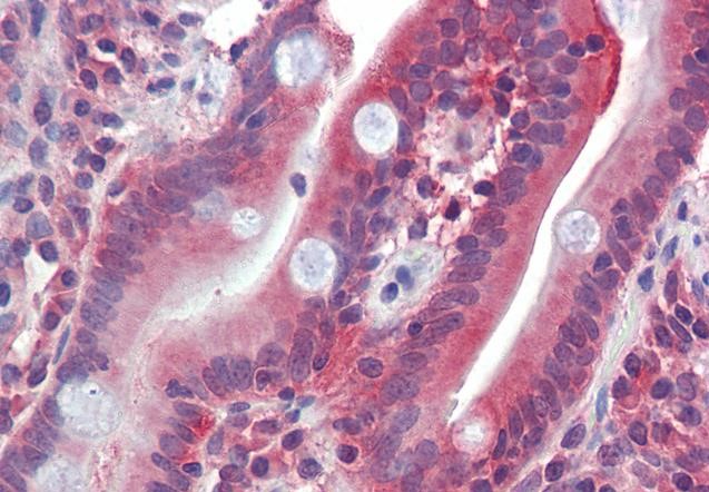
. SLC2A5 was detected in paraffin-embedded section of human intestinal cancer tissues. Heat mediated antigen retrieval was performed in citrate buffer (pH6, epitope retrieval solution) for 20 mins. The tissue section was blocked with 10% goat serum. The tissue section was then incubated with 1microg/ml rabbit anti-SLC2A5 Antibody (PB9960) overnight at 4°C. Biotinylated goat anti-rabbit IgG was used as secondary antibody and incubated for 30 minutes at 37°C. The tissue section was developed using Strepavidin-Biotin-Complex (SABC)(Catalog # SA1022) with DAB as the chromogen.")
. SLC2A5 was detected in immunocytochemical section of U20S cell. Enzyme antigen retrieval was performed using IHC enzyme antigen retrieval reagent (AR0022) for 15 mins. The cells were blocked with 10% goat serum. And then incubated with 2microg/mL rabbit anti-SLC2A5 Antibody (PB9960) overnight at 4°C. DyLight®488 Conjugated Goat Anti-Rabbit IgG (BA1127) was used as secondary antibody at 1:100 dilution and incubated for 30 minutes at 37°C. The section was counterstained with DAPI. Visualize using a fluorescence microscope and filter sets appropriate for the label used.")
. Overlay histogram showing THP-1 cells stained with PB9960 (Blue line). To facilitate intracellular staining, cells were fixed with 4% paraformaldehyde and permeabilized with permeabilization buffer. The cells were blocked with 10% normal goat serum. And then incubated with rabbit anti-SLC2A5 Antibody (PB9960,1microg/1x106 cells) for 30 min at 20°C. DyLight®488 conjugated goat anti-rabbit IgG (BA1127, 5-10microg/1x106 cells) was used as secondary antibody for 30 minutes at 20°C. Isotype control antibody (Green line) was rabbit IgG (1microg/1x106) used under the same conditions. Unlabelled sample without incubation with primary antibody and secondary antibody (Red line) was used as a blank control.")
Figure 1. Western blot analysis of SLC2A5 using anti-SLC2A5 antibody (PB9960). Electrophoresis was performed on a 5-20% SDS-PAGE gel at 70V (Stacking gel) / 90V (Resolving gel) for 2-3 hours. The sample well of each lane was loaded with 50ug of sample under reducing conditions. Lane 1: rat brain tissue lysates, Lane 2: K562 whole cell lysates. After Electrophoresis, proteins were transferred to a Nitrocellulose membrane at 150mA for 50-90 minutes. Blocked the membrane with 5% Non-fat Milk/ TBS for 1.5 hour at RT. The membrane was incubated with rabbit anti-SLC2A5 antigen affinity purified polyclonal antibody (Catalog # PB9960) at 0.5 microg/mL overnight at 4°C, then washed with TBS-0.1%Tween 3 times with 5 minutes each and probed with a goat anti-rabbit IgG-HRP secondary antibody at a dilution of 1:10000 for 1.5 hour at RT. The signal is developed using an Enhanced Chemiluminescent detection (ECL) kit (Catalog # EK1002) with Tanon 5200 system. A specific band was detected for SLC2A5 at approximately 55KD. The expected band size for SLC2A5 is at 55KD.
Anti-Glucose Transporter 5 GLUT5/SLC2A5 Antibody Picoband(r)
PB9960
ApplicationsFlow Cytometry, ImmunoFluorescence, Western Blot, ImmunoCytoChemistry, ImmunoHistoChemistry, ImmunoHistoChemistry Frozen
Product group Antibodies
ReactivityHuman, Rat
TargetSLC2A5
Overview
- SupplierBoster Bio
- Product NameAnti-Glucose Transporter 5 GLUT5/SLC2A5 Antibody Picoband(r)
- Delivery Days Customer9
- Application Supplier NoteTested Species: In-house tested species with positive results. By Heat: Boiling the paraffin sections in 10mM citrate buffer, pH6.0, for 20mins is required for the staining of formalin/paraffin sections. Other applications have not been tested. Optimal dilutions should be determined by end users.
- ApplicationsFlow Cytometry, ImmunoFluorescence, Western Blot, ImmunoCytoChemistry, ImmunoHistoChemistry, ImmunoHistoChemistry Frozen
- CertificationResearch Use Only
- ClonalityPolyclonal
- Concentration500 ug/ml
- Gene ID6518
- Target nameSLC2A5
- Target descriptionsolute carrier family 2 member 5
- Target synonymsGLUT-5, GLUT5, solute carrier family 2, facilitated glucose transporter member 5, glucose transporter type 5, small intestine, glucose transporter-like protein 5, solute carrier family 2 (facilitated glucose/fructose transporter), member 5, testicular tissue protein Li 81
- HostRabbit
- IsotypeIgG
- Protein IDP22732
- Protein NameSolute carrier family 2, facilitated glucose transporter member 5
- Scientific DescriptionBoster Bio Anti-Glucose Transporter 5 GLUT5/SLC2A5 Antibody Picoband® catalog # PB9960. Tested in Flow Cytometry, IF, IHC, IHC-F, ICC, WB applications. This antibody reacts with Human, Rat. The brand Picoband indicates this is a premium antibody that guarantees superior quality, high affinity, and strong signals with minimal background in Western blot applications. Only our best-performing antibodies are designated as Picoband, ensuring unmatched performance.
- ReactivityHuman, Rat
- Storage Instruction-20°C,2°C to 8°C
- UNSPSC12352203
References
- Yildirim OG, Guney C, Alcigir ME, et al. High-fructose consumption suppresses insulin signaling pathway accompanied by activation of macrophage and apoptotic markers in rat testis. Reprod Biol. 2023,23(4):100815. doi: 10.1016/j.repbio.2023.100815Read this paper
Datasheet
MSDS
Related products
Product group Antibodies
Anti-GLUT5 AntibodyA286017
ApplicationsELISA, ImmunoHistoChemistry
ReactivityHuman
- SizePrice
Product group Antibodies
Anti-SLC2A5 Antibody144-62851
ApplicationsWestern Blot
ReactivityHuman, Mouse
TargetSLC2A5
- SizePrice
Product group Antibodies

Goat anti-GLUT5 / SLC2A5EB09482
ApplicationsELISA, ImmunoHistoChemistry
ReactivityHuman
TargetSLC2A5
- SizePrice
Product group Antibodies
SLC2A5 AntibodyCSB-PA005624
ApplicationsWestern Blot, ELISA
ReactivityHuman
TargetSLC2A5
- SizePrice
Product group Antibodies

SLC2A5 / GLUT5 Antibody (N-Terminus)LS-C368951
ApplicationsWestern Blot
ReactivityHuman
TargetSLC2A5
- SizePrice
Product group Antibodies
Anti-SLC2A5 AntibodyHPA005449
ApplicationsWestern Blot, ImmunoHistoChemistry
ReactivityHuman
TargetSLC2A5
- SizePrice
Product group Antibodies
GLUT5 antibodyGTX47800
ApplicationsImmunoPrecipitation, Western Blot, ELISA, ImmunoHistoChemistry, ImmunoHistoChemistry Frozen, ImmunoHistoChemistry Paraffin
ReactivityHuman, Monkey
TargetSLC2A5
- SizePrice
Product group Antibodies
Anti-Glucose Transporter 5 GLUT5/SLC2A5 Antibody Picoband(r)PB9960-CARRIER-FREE
ApplicationsFlow Cytometry, ImmunoFluorescence, Western Blot, ImmunoCytoChemistry, ImmunoHistoChemistry, ImmunoHistoChemistry Frozen
ReactivityHuman, Rat
TargetSLC2A5
- SizePrice