


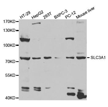
Figure 1. Western blot analysis of SLC3A1 using anti-SLC3A1 antibody (A02654-2). Electrophoresis was performed on a 5-20% SDS-PAGE gel at 70V (Stacking gel) / 90V (Resolving gel) for 2-3 hours. The sample well of each lane was loaded with 30 ug of sample under reducing conditions. Lane 1: monkey COS-7 whole cell lysates, Lane 2: human Caco-2 whole cell lysates, Lane 3: rat NRK whole cell lysates, Lane 4: mouse HBZY whole cell lysates. After electrophoresis, proteins were transferred to a nitrocellulose membrane at 150 mA for 50-90 minutes. Blocked the membrane with 5% non-fat milk/TBS for 1.5 hour at RT. The membrane was incubated with rabbit anti-SLC3A1 antigen affinity purified polyclonal antibody (Catalog # A02654-2) at 0.5 microg/mL overnight at 4°C, then washed with TBS-0.1%Tween 3 times with 5 minutes each and probed with a goat anti-rabbit IgG-HRP secondary antibody at a dilution of 1:5000 for 1.5 hour at RT. The signal is developed using an Enhanced Chemiluminescent detection (ECL) kit (Catalog # EK1002) with Tanon 5200 system. A specific band was detected for SLC3A1 at approximately 110 kDa. The expected band size for SLC3A1 is at 79 kDa.
. Overlay histogram showing U20S cells stained with A02654-2 (Blue line). The cells were fixed with 4% paraformaldehyde and blocked with 10% normal goat serum. And then incubated with rabbit anti-SLC3A1 Antibody (A02654-2, 1 microg/1x106 cells) for 30 min at 20°C. DyLight®488 conjugated goat anti-rabbit IgG (BA1127, 5-10 microg/1x106 cells) was used as secondary antibody for 30 minutes at 20°C. Isotype control antibody (Green line) was rabbit IgG (1 microg/1x106) used under the same conditions. Unlabelled sample without incubation with primary antibody and secondary antibody (Red line) was used as a blank control.")
Figure 1. Western blot analysis of SLC3A1 using anti-SLC3A1 antibody (A02654-2). Electrophoresis was performed on a 5-20% SDS-PAGE gel at 70V (Stacking gel) / 90V (Resolving gel) for 2-3 hours. The sample well of each lane was loaded with 30 ug of sample under reducing conditions. Lane 1: monkey COS-7 whole cell lysates, Lane 2: human Caco-2 whole cell lysates, Lane 3: rat NRK whole cell lysates, Lane 4: mouse HBZY whole cell lysates. After electrophoresis, proteins were transferred to a nitrocellulose membrane at 150 mA for 50-90 minutes. Blocked the membrane with 5% non-fat milk/TBS for 1.5 hour at RT. The membrane was incubated with rabbit anti-SLC3A1 antigen affinity purified polyclonal antibody (Catalog # A02654-2) at 0.5 microg/mL overnight at 4°C, then washed with TBS-0.1%Tween 3 times with 5 minutes each and probed with a goat anti-rabbit IgG-HRP secondary antibody at a dilution of 1:5000 for 1.5 hour at RT. The signal is developed using an Enhanced Chemiluminescent detection (ECL) kit (Catalog # EK1002) with Tanon 5200 system. A specific band was detected for SLC3A1 at approximately 110 kDa. The expected band size for SLC3A1 is at 79 kDa.
Anti-SLC3A1 Antibody Picoband(r)
A02654-2-CARRIER-FREE
ApplicationsFlow Cytometry, Western Blot, ELISA
Product group Antibodies
ReactivityHuman, Monkey, Mouse, Rat
TargetSLC3A1
Overview
- SupplierBoster Bio
- Product NameAnti-SLC3A1 Antibody Picoband(r)
- Delivery Days Customer9
- ApplicationsFlow Cytometry, Western Blot, ELISA
- CertificationResearch Use Only
- ClonalityPolyclonal
- Concentration500 ug/ml
- Gene ID6519
- Target nameSLC3A1
- Target descriptionsolute carrier family 3 member 1
- Target synonymsATR1, CSNU1, D2H, NBAT, RBAT, amino acid transporter heavy chain SLC3A1, B(0,+)-type amino acid transport protein, amino acid transporter 1, b(0,+)-type amino acid transporter-related heavy chain, neutral and basic amino acid transport protein rBAT, solute carrier family 3 (amino acid transporter heavy chain), member 1, solute carrier family 3 (cystine, dibasic and neutral amino acid transporters), member 1, solute carrier family 3 (cystine, dibasic and neutral amino acid transporters, activator of cystine, dibasic and neutral amino acid transport), member 1
- HostRabbit
- IsotypeIgG
- Protein IDQ07837
- Protein NameAmino acid transporter heavy chain SLC3A1
- Scientific DescriptionBoster Bio Anti-SLC3A1 Antibody Picoband® catalog # A02654-2. Tested in ELISA, Flow Cytometry, WB applications. This antibody reacts with Human, Monkey, Mouse, Rat. The brand Picoband indicates this is a premium antibody that guarantees superior quality, high affinity, and strong signals with minimal background in Western blot applications. Only our best-performing antibodies are designated as Picoband, ensuring unmatched performance.
- ReactivityHuman, Monkey, Mouse, Rat
- Storage Instruction-20°C,2°C to 8°C
- UNSPSC12352203
Related products
Product group Antibodies
Anti-SLC3A1 AntibodyA30808
ApplicationsImmunoFluorescence, Western Blot, ImmunoHistoChemistry
ReactivityHuman, Mouse, Rat
- SizePrice
Product group Antibodies
Anti-SLC3A1 Antibody144-05500
ApplicationsImmunoFluorescence, Western Blot
ReactivityHuman, Mouse
TargetSLC3A1
- SizePrice
Product group Antibodies
SLC3A1 AntibodyCSB-PA737352LA01HU
ApplicationsImmunoFluorescence, Western Blot, ELISA, ImmunoHistoChemistry
ReactivityHuman, Mouse, Rat
TargetSLC3A1
- SizePrice
Product group Antibodies
SLC3A1 Polyclonal AntibodyCAC14903
ApplicationsImmunoFluorescence, Western Blot, ELISA, ImmunoHistoChemistry
ReactivityMouse, Rat
TargetSLC3A1
- SizePrice
Product group Antibodies

SLC3A1 / ATR1 AntibodyLS-C334095
ApplicationsImmunoFluorescence, Western Blot, ImmunoHistoChemistry
ReactivityHuman, Mouse
TargetSLC3A1
- SizePrice
Product group Antibodies
SLC3A1 antibodyGTX33506
ApplicationsImmunoFluorescence, Western Blot, ImmunoCytoChemistry, ImmunoHistoChemistry, ImmunoHistoChemistry Paraffin
ReactivityHuman, Mouse, Rat
TargetSLC3A1
- SizePrice

Product group Antibodies
Anti-SLC3A1-25ulHPA038360
ApplicationsWestern Blot, ImmunoHistoChemistry
ReactivityHuman
- SizePrice
Product group Antibodies
Anti-SLC3A1 AntibodyCAB5500
ApplicationsWestern Blot, ELISA, ImmunoHistoChemistry, ImmunoHistoChemistry Paraffin
ReactivityHuman
TargetSLC3A1
- SizePrice