


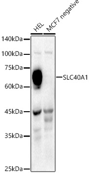
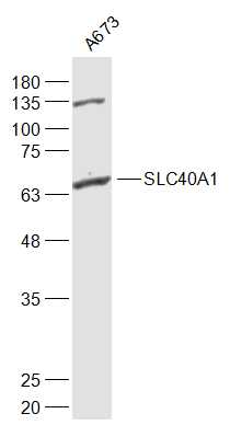
Figure 1. Western blot analysis of SLC40A1 using anti-SLC40A1 antibody (A01953-2). Electrophoresis was performed on a 5-20% SDS-PAGE gel at 70V (Stacking gel) / 90V (Resolving gel) for 2-3 hours. The sample well of each lane was loaded with 30 ug of sample unde r reducing conditions. Lane 1: human placenta tissue lysates, Lane 2: rat liver tissue lysates, Lane 3: mouse spleen tissue lysates, Lane 4: mouse liver tissue lysates. After electrophoresis, proteins were transferred to a nitrocellulose membrane at 150 mA for 50-90 minutes. Blocked the membrane with 5% non-fat milk/TBS for 1.5 hour at RT. The membrane was incubated with rabbit anti-SLC40A1 antigen affinity purified polyclonal antibody (Catalog # A01953-2) at 0.5 microg/mL overnight at 4°C, then washed with TBS-0.1%Tween 3 times with 5 minutes each and probed with a goat anti-rabbit IgG-HRP secondary antibody at a dilution of 1:5000 for 1.5 hour at RT. The signal is developed using an Enhanced Chemiluminescent detection (ECL) kit (Catalog # EK1002) with Tanon 5200 system. A specific band was detected for SLC40A1 at approximately 70 kDa. The expected band size for SLC40A1 is at 63 kDa.
. SLC40A1 was detected in a paraffin-embedded section of human larynx squamous cell carcinoma tissue. Heat mediated antigen retrieval was performed in EDTA buffer (pH 8.0, epitope retrieval solution). The tissue section was blocked with 10% goat serum. The tissue section was then incubated with 2 microg/ml rabbit anti-SLC40A1 Antibody (A01953-2) overnight at 4°C. Peroxidase Conjugated Goat Anti-rabbit IgG was used as secondary antibody and incubated for 30 minutes at 37°C. The tissue section was developed using HRP Conjugated Rabbit IgG Super Vision Assay Kit (Catalog # SV0002) with DAB as the chromogen.")
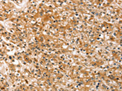
. SLC40A1 was detected in a paraffin-embedded section of human liver cancer tissue. Heat mediated antigen retrieval was performed in EDTA buffer (pH 8.0, epitope retrieval solution). The tissue section was blocked with 10% goat serum. The tissue section was then incubated with 2 microg/ml rabbit anti-SLC40A1 Antibody (A01953-2) overnight at 4°C. Peroxidase Conjugated Goat Anti-rabbit IgG was used as secondary antibody and incubated for 30 minutes at 37°C. The tissue section was developed using HRP Conjugated Rabbit IgG Super Vision Assay Kit (Catalog # SV0002) with DAB as the chromogen.")
. SLC40A1 was detected in a paraffin-embedded section of human ovarian serous adenocarcinoma tissue. Heat mediated antigen retrieval was performed in EDTA buffer (pH 8.0, epitope retrieval solution). The tissue section was blocked with 10% goat serum. The tissue section was then incubated with 2 microg/ml rabbit anti-SLC40A1 Antibody (A01953-2) overnight at 4°C. Peroxidase Conjugated Goat Anti-rabbit IgG was used as secondary antibody and incubated for 30 minutes at 37°C. The tissue section was developed using HRP Conjugated Rabbit IgG Super Vision Assay Kit (Catalog # SV0002) with DAB as the chromogen.")
. SLC40A1 was detected in a paraffin-embedded section of human prostate adenocarcinoma tissue. Heat mediated antigen retrieval was performed in EDTA buffer (pH 8.0, epitope retrieval solution). The tissue section was blocked with 10% goat serum. The tissue section was then incubated with 2 microg/ml rabbit anti-SLC40A1 Antibody (A01953-2) overnight at 4°C. Peroxidase Conjugated Goat Anti-rabbit IgG was used as secondary antibody and incubated for 30 minutes at 37°C. The tissue section was developed using HRP Conjugated Rabbit IgG Super Vision Assay Kit (Catalog # SV0002) with DAB as the chromogen.")
. SLC40A1 was detected in a paraffin-embedded section of human rectum adenocarcinoma tissue. Heat mediated antigen retrieval was performed in EDTA buffer (pH 8.0, epitope retrieval solution). The tissue section was blocked with 10% goat serum. The tissue section was then incubated with 2 microg/ml rabbit anti-SLC40A1 Antibody (A01953-2) overnight at 4°C. Peroxidase Conjugated Goat Anti-rabbit IgG was used as secondary antibody and incubated for 30 minutes at 37°C. The tissue section was developed using HRP Conjugated Rabbit IgG Super Vision Assay Kit (Catalog # SV0002) with DAB as the chromogen.")
. SLC40A1 was detected in a paraffin-embedded section of human ovarian cancer tissue. Heat mediated antigen retrieval was performed in EDTA buffer (pH 8.0, epitope retrieval solution). The tissue section was blocked with 10% goat serum. The tissue section was then incubated with 5 microg/mL rabbit anti-SLC40A1 Antibody (A01953-2) overnight at 4°C. FITC Conjugated Goat Anti-Rabbit IgG (BA1105) was used as secondary antibody at 1:500 dilution and incubated for 30 minutes at 37°C. The section was counterstained with DAPI. Visualize using a fluorescence microscope and filter sets appropriate for the label used.")
. Overlay histogram showing U20S cells stained with A01953-2 (Blue line). To facilitate intracellular staining, cells were fixed with 4% paraformaldehyde and permeabilized with permeabilization buffer. The cells were blocked with 10% normal goat serum. And then incubated with rabbit anti-SLC40A1 Antibody (A01953-2, 1 microg/1x106 cells) for 30 min at 20°C. DyLight®488 conjugated goat anti-rabbit IgG (BA1127, 5-10 microg/1x106 cells) was used as secondary antibody for 30 minutes at 20°C. Isotype control antibody (Green line) was rabbit IgG (1 microg/1x106) used under the same conditions. Unlabelled sample without incubation with primary antibody and secondary antibody (Red line) was used as a blank control.")
Figure 1. Western blot analysis of SLC40A1 using anti-SLC40A1 antibody (A01953-2). Electrophoresis was performed on a 5-20% SDS-PAGE gel at 70V (Stacking gel) / 90V (Resolving gel) for 2-3 hours. The sample well of each lane was loaded with 30 ug of sample unde r reducing conditions. Lane 1: human placenta tissue lysates, Lane 2: rat liver tissue lysates, Lane 3: mouse spleen tissue lysates, Lane 4: mouse liver tissue lysates. After electrophoresis, proteins were transferred to a nitrocellulose membrane at 150 mA for 50-90 minutes. Blocked the membrane with 5% non-fat milk/TBS for 1.5 hour at RT. The membrane was incubated with rabbit anti-SLC40A1 antigen affinity purified polyclonal antibody (Catalog # A01953-2) at 0.5 microg/mL overnight at 4°C, then washed with TBS-0.1%Tween 3 times with 5 minutes each and probed with a goat anti-rabbit IgG-HRP secondary antibody at a dilution of 1:5000 for 1.5 hour at RT. The signal is developed using an Enhanced Chemiluminescent detection (ECL) kit (Catalog # EK1002) with Tanon 5200 system. A specific band was detected for SLC40A1 at approximately 70 kDa. The expected band size for SLC40A1 is at 63 kDa.
Anti-SLC40A1 Antibody Picoband(r)
A01953-2-CARRIER-FREE
ApplicationsFlow Cytometry, ImmunoFluorescence, Western Blot, ELISA, ImmunoHistoChemistry
Product group Antibodies
ReactivityHuman, Mouse, Rat
TargetSLC40A1
Overview
- SupplierBoster Bio
- Product NameAnti-SLC40A1 Antibody Picoband(r)
- Delivery Days Customer9
- ApplicationsFlow Cytometry, ImmunoFluorescence, Western Blot, ELISA, ImmunoHistoChemistry
- CertificationResearch Use Only
- ClonalityPolyclonal
- Concentration500 ug/ml
- Gene ID30061
- Target nameSLC40A1
- Target descriptionsolute carrier family 40 member 1
- Target synonymsFPN, FPN1, HFE4, IREG1, MST079, MSTP079, MTP1, SLC11A3, ferroportin, SLC40 iron transporter, iron regulated gene 1, solute carrier family 11 (proton-coupled divalent metal ion transporters), member 3, solute carrier family 40 (iron-regulated transporter), member 1
- HostRabbit
- IsotypeIgG
- Protein IDQ9NP59
- Protein NameFerroportin
- Scientific DescriptionBoster Bio Anti-SLC40A1 Antibody Picoband® catalog # A01953-2. Tested in ELISA, Flow Cytometry, IF, IHC, WB applications. This antibody reacts with Human, Mouse, Rat. The brand Picoband indicates this is a premium antibody that guarantees superior quality, high affinity, and strong signals with minimal background in Western blot applications. Only our best-performing antibodies are designated as Picoband, ensuring unmatched performance.
- ReactivityHuman, Mouse, Rat
- Storage Instruction-20°C,2°C to 8°C
- UNSPSC12352203
Related products
Product group Antibodies
Anti-SLC40A1 AntibodyA91593
ApplicationsWestern Blot
ReactivityHuman, Mouse
- SizePrice
Product group Antibodies
ApplicationsWestern Blot, ELISA
ReactivityHuman, Mouse
TargetSLC40A1
- SizePrice
Product group Antibodies

SLC40A1 / Ferroportin-1 AntibodyLS-C830077
ApplicationsELISA, ImmunoHistoChemistry
ReactivityHuman
TargetSLC40A1
- SizePrice
Product group Antibodies
References
SLC40A1 Polyclonal AntibodyBS-4906R
ApplicationsWestern Blot, ELISA, ImmunoHistoChemistry, ImmunoHistoChemistry Paraffin
ReactivityBovine, Canine, Human, Mouse, Porcine, Rabbit, Rat
TargetSLC40A1
- SizePrice
Product group Antibodies

ApplicationsFlow Cytometry, ImmunoFluorescence, ELISA
ReactivityHuman
TargetSLC40A1
- SizePrice
Product group Antibodies
SLC40A1 AntibodyCSB-PA572147
ApplicationsELISA, ImmunoHistoChemistry
ReactivityHuman
TargetSLC40A1
- SizePrice
Product group Antibodies
ApplicationsImmunoPrecipitation, Western Blot, ImmunoCytoChemistry, ImmunoHistoChemistry
ReactivityMouse, Porcine, Rat
TargetSLC40A1
- SizePrice
Product group Antibodies
Ferroportin 1 antibodyGTX85744
ApplicationsWestern Blot, ELISA, ImmunoHistoChemistry, ImmunoHistoChemistry Paraffin
ReactivityHuman, Mouse
TargetSLC40A1
- SizePrice

Product group Antibodies
Anti-SLC40A1 AntibodyHPA065634
ApplicationsImmunoCytoChemistry, ImmunoHistoChemistry
ReactivityHuman
TargetSLC40A1
- SizePrice