

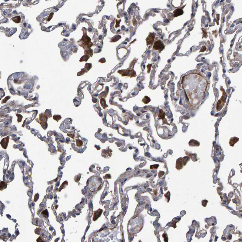
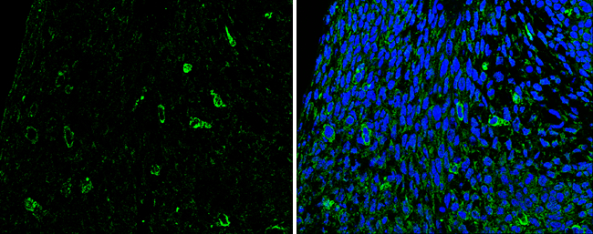
Immunohistochemical staining of human lung shows strong cytoplasmic positivity in macrophages.
Immunohistochemical staining of human lung shows strong cytoplasmic positivity in macrophages.
Anti-SLIT2 Antibody
HPA023088
ApplicationsImmunoHistoChemistry
Product group Antibodies
ReactivityHuman
TargetSLIT2
Overview
- SupplierAtlas Antibodies
- Product NameAnti-SLIT2 Antibody
- Delivery Days Customer4
- ApplicationsImmunoHistoChemistry
- CertificationResearch Use Only
- ClonalityPolyclonal
- ConjugateUnconjugated
- Gene ID9353
- Target nameSLIT2
- Target descriptionslit guidance ligand 2
- Target synonymsSLIL3, Slit-2, slit homolog 2 protein
- HostRabbit
- IsotypeIgG
- Protein IDO94813
- Protein NameSlit homolog 2 protein
- Scientific DescriptionRecombinant Protein Epitope Signature Tag (PrEST) antigen sequence
- ReactivityHuman
- Storage Instruction-20°C,2°C to 8°C
- UNSPSC41116161
Datasheet
MSDS
Related products
Product group Antibodies
Anti-SLIT2 AntibodyA46292
ApplicationsImmunoHistoChemistry
ReactivityHuman, Mouse
- SizePrice

Product group Antibodies
Anti-SLIT2 Antibody Picoband(r)A01627-1-CARRIER-FREE
ApplicationsFlow Cytometry, Western Blot, ELISA
ReactivityHuman
TargetSLIT2
- SizePrice
Product group Antibodies
SLIT2 Polyclonal AntibodyBS-2743R
ApplicationsImmunoFluorescence, Western Blot, ELISA, ImmunoCytoChemistry, ImmunoHistoChemistry, ImmunoHistoChemistry Frozen, ImmunoHistoChemistry Paraffin
ReactivityHuman, Mouse, Rat
TargetSLIT2
- SizePrice
Product group Antibodies
SLIT2 AntibodyCSB-PA136864
ApplicationsWestern Blot, ELISA, ImmunoHistoChemistry
ReactivityHuman, Mouse, Rat
TargetSLIT2
- SizePrice
Product group Antibodies

Goat anti-SLIT2EB07287
ApplicationsELISA, ImmunoHistoChemistry
ReactivityCanine, Human, Mouse, Rat
TargetSLIT2
- SizePrice
Product group Antibodies
ApplicationsImmunoPrecipitation, Western Blot, ImmunoCytoChemistry, ImmunoHistoChemistry
ReactivityMouse, Rat
TargetSLIT2
- SizePrice
Product group Antibodies

SLIT2 AntibodyLS-C403581
ApplicationsWestern Blot, ELISA, ImmunoHistoChemistry
ReactivityHuman, Mouse, Rat
TargetSLIT2
- SizePrice
Product group Antibodies
SLIT2 antibodyGTX118220
ApplicationsWestern Blot, ImmunoHistoChemistry, ImmunoHistoChemistry Paraffin
ReactivityHuman, Mouse, Rat
TargetSLIT2
- SizePrice