


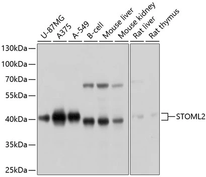
Figure 1. Western blot analysis of STOML2 using anti-STOML2 antibody (M04108-1). Electrophoresis was performed on a 5-20% SDS-PAGE gel at 70V (Stacking gel) / 90V (Resolving gel) for 2-3 hours. The sample well of each lane was loaded with 30 ug of sample under reducing conditions. Lane 1: human Jurkat whole cell lysates, Lane 2: human Hela whole cell lysates, Lane 3: human Caco-2 whole cell lysates, Lane 4: human A549 whole cell lysates, Lane 5: rat brain tissue lysates, Lane 6: rat C6 whole cell lysates, Lane 7: mouse brain tissue lysates, Lane 8: mouse RAW264.7 whole cell lysates. After electrophoresis, proteins were transferred to a nitrocellulose membrane at 150 mA for 50-90 minutes. Blocked the membrane with 5% non-fat milk/TBS for 1.5 hour at RT. The membrane was incubated with rabbit anti-STOML2 antigen affinity purified monoclonal antibody (M04108-1) at 1:500 overnight at 4°C, then washed with TBS-0.1%Tween 3 times with 5 minutes each and probed with a goat anti-rabbit IgG-HRP secondary antibody at a dilution of 1:500 for 1.5 hour at RT. The signal is developed using an Enhanced Chemiluminescent detection (ECL) kit (Catalog # EK1002) with Tanon 5200 system. A specific band was detected for STOML2 at approximately 39 kDa. The expected band size for STOML2 is at 39 kDa.
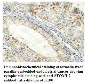
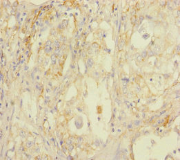
 analysis using the Antibody at 1:50 dilution. (wb at 1:1K dilution)")
Figure 1. Western blot analysis of STOML2 using anti-STOML2 antibody (M04108-1). Electrophoresis was performed on a 5-20% SDS-PAGE gel at 70V (Stacking gel) / 90V (Resolving gel) for 2-3 hours. The sample well of each lane was loaded with 30 ug of sample under reducing conditions. Lane 1: human Jurkat whole cell lysates, Lane 2: human Hela whole cell lysates, Lane 3: human Caco-2 whole cell lysates, Lane 4: human A549 whole cell lysates, Lane 5: rat brain tissue lysates, Lane 6: rat C6 whole cell lysates, Lane 7: mouse brain tissue lysates, Lane 8: mouse RAW264.7 whole cell lysates. After electrophoresis, proteins were transferred to a nitrocellulose membrane at 150 mA for 50-90 minutes. Blocked the membrane with 5% non-fat milk/TBS for 1.5 hour at RT. The membrane was incubated with rabbit anti-STOML2 antigen affinity purified monoclonal antibody (M04108-1) at 1:500 overnight at 4°C, then washed with TBS-0.1%Tween 3 times with 5 minutes each and probed with a goat anti-rabbit IgG-HRP secondary antibody at a dilution of 1:500 for 1.5 hour at RT. The signal is developed using an Enhanced Chemiluminescent detection (ECL) kit (Catalog # EK1002) with Tanon 5200 system. A specific band was detected for STOML2 at approximately 39 kDa. The expected band size for STOML2 is at 39 kDa.
Anti-SLP2 Rabbit Monoclonal Antibody
M04108-1
ApplicationsFlow Cytometry, ImmunoFluorescence, ImmunoPrecipitation, Western Blot, ImmunoCytoChemistry
Product group Antibodies
ReactivityHuman, Mouse, Rat
TargetSTOML2
Overview
- SupplierBoster Bio
- Product NameAnti-SLP2 Rabbit Monoclonal Antibody
- Delivery Days Customer9
- ApplicationsFlow Cytometry, ImmunoFluorescence, ImmunoPrecipitation, Western Blot, ImmunoCytoChemistry
- CertificationResearch Use Only
- ClonalityMonoclonal
- Clone ID28S56
- Gene ID30968
- Target nameSTOML2
- Target descriptionstomatin like 2
- Target synonymsHSPC108, SLP-2, stomatin-like protein 2, mitochondrial, EPB72-like 2, EPB72-like protein 2, paraprotein target 7, paratarg-7, stomatin (EPB72)-like 2
- HostRabbit
- IsotypeIgG
- Protein IDQ9UJZ1
- Protein NameStomatin-like protein 2, mitochondrial
- Scientific DescriptionBoster Bio Anti-SLP2 Rabbit Monoclonal Antibody catalog # M04108-1. Tested in WB, ICC/IF, IP, Flow Cytometry applications. This antibody reacts with Human, Mouse, Rat.
- ReactivityHuman, Mouse, Rat
- Storage Instruction-20°C
- UNSPSC12352203
Related products
Product group Antibodies
Anti-STOML2 AntibodyA47820
ApplicationsWestern Blot, ELISA, ImmunoHistoChemistry
ReactivityHuman, Mouse, Rat
- SizePrice
Product group Antibodies
Anti-STOML2 Antibody144-10398
ApplicationsWestern Blot
ReactivityHuman, Mouse, Rat
TargetSTOML2
- SizePrice
Product group Antibodies
ApplicationsFlow Cytometry, Western Blot, ImmunoCytoChemistry
ReactivityHuman, Mouse, Rat
TargetSTOML2
- SizePrice
Product group Antibodies
STOML2 AntibodyCSB-PA892131ESR1HU
ApplicationsELISA, ImmunoHistoChemistry
ReactivityHuman
TargetSTOML2
- SizePrice

Product group Antibodies
Anti-STOML2 AntibodyHPA062016
ApplicationsWestern Blot, ImmunoHistoChemistry
ReactivityHuman
TargetSTOML2
- SizePrice
Product group Antibodies
STOML2 antibodyGTX64801
ApplicationsWestern Blot, ImmunoHistoChemistry, ImmunoHistoChemistry Paraffin
ReactivityHuman, Mouse
TargetSTOML2
- SizePrice
Product group Antibodies

STOML2 AntibodyLS-C763461
ApplicationsImmunoHistoChemistry
ReactivityHuman
TargetSTOML2
- SizePrice
Product group Antibodies
Anti-STOML2Y158357
ApplicationsWestern Blot, ELISA, ImmunoHistoChemistry
ReactivityHuman
- SizePrice