


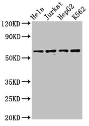
Figure 1. Western blot analysis of SMARCD1 using anti-SMARCD1 antibody (A07318-1). Electrophoresis was performed on a 5-20% SDS-PAGE gel at 70V (Stacking gel) / 90V (Resolving gel) for 2-3 hours. The sample well of each lane was loaded with 30 ug of sample under reducing conditions. Lane 1: human Hela whole cell lysates, Lane 2: human 293T whole cell lysates, Lane 3: human PANC-1 whole cell lysates, Lane 4: human Jurkat whole cell lysates, Lane 5: human MOLT-4 whole cell lysates, Lane 6: human Caco-2 whole cell lysates, Lane 7: human SH-SY5Y whole cell lysates, Lane 8: human Daudi whole cell lysates. After electrophoresis, proteins were transferred to a nitrocellulose membrane at 150 mA for 50-90 minutes. Blocked the membrane with 5% non-fat milk/TBS for 1.5 hour at RT. The membrane was incubated with rabbit anti-SMARCD1 antigen affinity purified polyclonal antibody (Catalog # A07318-1) at 0.5 microg/mL overnight at 4°C, then washed with TBS-0.1%Tween 3 times with 5 minutes each and probed with a goat anti-rabbit IgG-HRP secondary antibody at a dilution of 1:5000 for 1.5 hour at RT. The signal is developed using an Enhanced Chemiluminescent detection (ECL) kit (Catalog # EK1002) with Tanon 5200 system. A specific band was detected for SMARCD1 at approximately 58 kDa. The expected band size for SMARCD1 is at 58 kDa.
Figure 1. Western blot analysis of SMARCD1 using anti-SMARCD1 antibody (A07318-1). Electrophoresis was performed on a 5-20% SDS-PAGE gel at 70V (Stacking gel) / 90V (Resolving gel) for 2-3 hours. The sample well of each lane was loaded with 30 ug of sample under reducing conditions. Lane 1: human Hela whole cell lysates, Lane 2: human 293T whole cell lysates, Lane 3: human PANC-1 whole cell lysates, Lane 4: human Jurkat whole cell lysates, Lane 5: human MOLT-4 whole cell lysates, Lane 6: human Caco-2 whole cell lysates, Lane 7: human SH-SY5Y whole cell lysates, Lane 8: human Daudi whole cell lysates. After electrophoresis, proteins were transferred to a nitrocellulose membrane at 150 mA for 50-90 minutes. Blocked the membrane with 5% non-fat milk/TBS for 1.5 hour at RT. The membrane was incubated with rabbit anti-SMARCD1 antigen affinity purified polyclonal antibody (Catalog # A07318-1) at 0.5 microg/mL overnight at 4°C, then washed with TBS-0.1%Tween 3 times with 5 minutes each and probed with a goat anti-rabbit IgG-HRP secondary antibody at a dilution of 1:5000 for 1.5 hour at RT. The signal is developed using an Enhanced Chemiluminescent detection (ECL) kit (Catalog # EK1002) with Tanon 5200 system. A specific band was detected for SMARCD1 at approximately 58 kDa. The expected band size for SMARCD1 is at 58 kDa.
Anti-SMARCD1 Antibody Picoband(r)
A07318-1-CARRIER-FREE
ApplicationsWestern Blot
Product group Antibodies
ReactivityHuman
TargetSMARCD1
Overview
- SupplierBoster Bio
- Product NameAnti-SMARCD1 Antibody Picoband(r)
- Delivery Days Customer9
- ApplicationsWestern Blot
- CertificationResearch Use Only
- ClonalityPolyclonal
- Concentration500 ug/ml
- Gene ID6602
- Target nameSMARCD1
- Target descriptionSWI/SNF related BAF chromatin remodeling complex subunit D1
- Target synonymsBAF60A, CRACD1, CSS11, Rsc6p, SWI/SNF-related matrix-associated actin-dependent regulator of chromatin subfamily D member 1, 60 kDa BRG-1/Brm-associated factor subunit A, BRG1-associated factor 60A, SWI/SNF complex 60 kDa subunit A, SWI/SNF related, matrix associated, actin dependent regulator of chromatin, subfamily d, member 1, Swp73-like protein, chromatin remodeling complex BAF60A subunit, mammalian chromatin remodeling complex BRG1-associated factor 60A
- HostRabbit
- IsotypeIgG
- Protein IDQ96GM5
- Protein NameSWI/SNF-related matrix-associated actin-dependent regulator of chromatin subfamily D member 1
- Scientific DescriptionBoster Bio Anti-SMARCD1 Antibody Picoband® catalog # A07318-1. Tested in WB applications. This antibody reacts with Human. The brand Picoband indicates this is a premium antibody that guarantees superior quality, high affinity, and strong signals with minimal background in Western blot applications. Only our best-performing antibodies are designated as Picoband, ensuring unmatched performance.
- ReactivityHuman
- Storage Instruction-20°C,2°C to 8°C
- UNSPSC12352203
Related products
Product group Antibodies
Anti-SMARCD1 AntibodyA31286
ApplicationsWestern Blot, ImmunoHistoChemistry
ReactivityHuman, Mouse, Rat
- SizePrice

Product group Antibodies
Anti-SMARCD1 AntibodyHPA004101
ApplicationsWestern Blot, ChIP Chromatin ImmunoPrecipitation, ImmunoCytoChemistry, ImmunoHistoChemistry
ReactivityHuman, Mouse, Rat
TargetSMARCD1
- SizePrice
Product group Antibodies
SMARCD1 AntibodyCSB-PA846627LA01HU
ApplicationsImmunoFluorescence, Western Blot, ELISA, ImmunoHistoChemistry
ReactivityHuman
TargetSMARCD1
- SizePrice
Product group Antibodies

SMARCD1 / BAF60A AntibodyLS-C334632
ApplicationsWestern Blot, ImmunoHistoChemistry
ReactivityHuman, Mouse, Rat
TargetSMARCD1
- SizePrice
Product group Antibodies
SMARCD1 Polyclonal AntibodyCAC15000
ApplicationsImmunoFluorescence, Western Blot, ELISA, ImmunoHistoChemistry
TargetSMARCD1
- SizePrice
Product group Antibodies
SMARCD1 Recombinant AntibodyBSM-60692R
ApplicationsWestern Blot
ReactivityHuman, Mouse
TargetSMARCD1
- SizePrice
![HeLa whole cell and nuclear extracts (30 μg) were separated by 10% SDS-PAGE, and the membrane was blotted with SMARCD1 antibody [N2C1], Internal (GTX114780) diluted at 1:1000.](https://www.genetex.com/upload/website/prouct_img/normal/GTX114780/GTX114780_40233_20161013_WB_Fraction_w_23060518_678.webp)
Product group Antibodies
SMARCD1 antibody [N2C1], InternalGTX114780
ApplicationsImmunoFluorescence, Western Blot, ImmunoCytoChemistry
ReactivityHuman, Mouse
TargetSMARCD1
- SizePrice
Product group Antibodies
Anti-SMARCD1 Antibody144-06310
ApplicationsWestern Blot
ReactivityHuman, Mouse, Rat
TargetSMARCD1
- SizePrice