

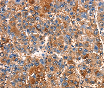

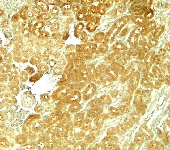
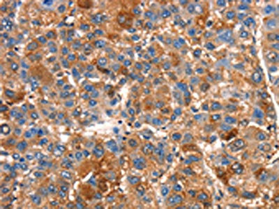
Anti-SMURF1 Antibody
A45516
ApplicationsImmunoHistoChemistry
Product group Antibodies
ReactivityHuman
Overview
- SupplierAntibodies.com
- Product NameAnti-SMURF1 Antibody
- Delivery Days Customer7
- ApplicationsImmunoHistoChemistry
- CertificationResearch Use Only
- ClonalityPolyclonal
- Concentration1.8 mg/ml
- ConjugateUnconjugated
- HostRabbit
- Scientific DescriptionRabbit polyclonal antibody to SMURF1
- ReactivityHuman
- UNSPSC12352203
Related products
Product group Antibodies

Anti-SMURF1 [RAB-C449]Ab01883-1.1
ApplicationsFlow Cytometry, ImmunoFluorescence
ReactivityHuman
TargetSMURF1
- SizePrice
Product group Antibodies
Anti-SMURF1 Antibody144-64492
ApplicationsWestern Blot
ReactivityHuman
TargetSMURF1
- SizePrice
Product group Antibodies
Smurf1 Polyclonal AntibodyCAC07256
ApplicationsImmunoFluorescence, Western Blot, ELISA, ImmunoHistoChemistry
ReactivityMouse
TargetSMURF1
- SizePrice

Product group Antibodies
Anti-SMURF1 Antibody Picoband(r)PB9892-CARRIER-FREE
ApplicationsFlow Cytometry, ImmunoFluorescence, Western Blot, ImmunoCytoChemistry, ImmunoHistoChemistry
ReactivityHamster, Human, Rat
TargetSMURF1
- SizePrice
Product group Antibodies
SMURF1 antibodyGTX17223
ApplicationsWestern Blot, ELISA, ImmunoHistoChemistry, ImmunoHistoChemistry Paraffin
ReactivityHuman, Mouse, Rat
TargetSMURF1
- SizePrice
Product group Antibodies
SMURF1 AntibodyCSB-PA026284
ApplicationsELISA, ImmunoHistoChemistry
ReactivityHuman, Mouse
TargetSMURF1
- SizePrice
Product group Antibodies

SMURF1 Antibody (HRP)LS-C501877
ApplicationsELISA
ReactivityHuman
TargetSMURF1
- SizePrice

Product group Antibodies
Anti-SMURF1 AntibodyHPA019671
ApplicationsChIP Chromatin ImmunoPrecipitation, ImmunoCytoChemistry
ReactivityHuman
TargetSMURF1
- SizePrice