


Immunohistochemical staining of human breast shows moderate cytoplasmic positivity in glandular cells.

Immunohistochemical staining of human breast shows moderate cytoplasmic positivity in glandular cells.
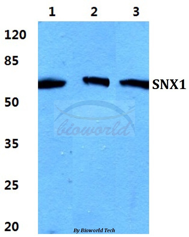
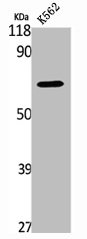
Anti-SNX1 Antibody
HPA047373
ApplicationsWestern Blot, ImmunoCytoChemistry, ImmunoHistoChemistry
Product group Antibodies
ReactivityHuman
TargetSNX1
Overview
- SupplierAtlas Antibodies
- Product NameAnti-SNX1 Antibody
- Delivery Days Customer4
- ApplicationsWestern Blot, ImmunoCytoChemistry, ImmunoHistoChemistry
- CertificationResearch Use Only
- ClonalityPolyclonal
- ConjugateUnconjugated
- Gene ID6642
- Target nameSNX1
- Target descriptionsorting nexin 1
- Target synonymsHsT17379, VPS5, sorting nexin-1, sorting nexin 1A
- HostRabbit
- IsotypeIgG
- Protein IDQ13596
- Protein NameSorting nexin-1
- Scientific DescriptionRecombinant Protein Epitope Signature Tag (PrEST) antigen sequence
- ReactivityHuman
- Storage Instruction-20°C,2°C to 8°C
- UNSPSC41116161
Datasheet
MSDS
Related products
Product group Antibodies
Anti-SNX1 AntibodyA28170
ApplicationsWestern Blot, ImmunoCytoChemistry
ReactivityHuman, Mouse, Rat
- SizePrice

Product group Antibodies
Anti-SNX1 Antibody Picoband(r)A02692-2-CARRIER-FREE
ApplicationsFlow Cytometry, ImmunoFluorescence, Western Blot, ELISA, ImmunoCytoChemistry, ImmunoHistoChemistry
ReactivityHuman
TargetSNX1
- SizePrice
Product group Antibodies
Anti-SNX1 Antibody144-08625
ApplicationsWestern Blot, ImmunoHistoChemistry
ReactivityHuman, Mouse, Rat
TargetSNX1
- SizePrice
Product group Antibodies
SNX1 Recombinant AntibodyBSM-61938R
ApplicationsFlow Cytometry, ImmunoFluorescence, Western Blot, ImmunoCytoChemistry, ImmunoHistoChemistry, ImmunoHistoChemistry Frozen, ImmunoHistoChemistry Paraffin
ReactivityHuman, Mouse, Rat
TargetSNX1
- SizePrice
Product group Antibodies
SNX1 AntibodyCSB-PA004128
ApplicationsWestern Blot, ELISA
ReactivityHuman, Mouse, Rat
TargetSNX1
- SizePrice
Product group Antibodies

Goat anti-SNX1EB09792
ApplicationsWestern Blot, ELISA, ImmunoCytoChemistry, ImmunoHistoChemistry
ReactivityBovine, Canine, Human, Mouse, Rat
TargetSNX1
- SizePrice
Product group Antibodies

SNX1 AntibodyLS-C410158
ApplicationsWestern Blot
ReactivityHuman, Mouse, Rat
TargetSNX1
- SizePrice

Product group Antibodies
Anti-SNX1 AntibodyHPA052761
ApplicationsWestern Blot, ImmunoCytoChemistry
ReactivityHuman
TargetSNX1
- SizePrice