


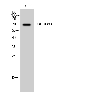
Figure 1. Western blot analysis of Spindly/SPDL1 using anti-Spindly/SPDL1 antibody (A12722-2). Electrophoresis was performed on a 5-20% SDS-PAGE gel at 70V (Stacking gel) / 90V (Resolving gel) for 2-3 hours. The sample well of each lane was loaded with 30 ug of sample under reducing conditions. Lane 1: human U251 whole cell lysates, Lane 2: human Hela whole cell lysates, Lane 3: human Caco-2 whole cell lysates, Lane 4: rat liver tissue lysates, Lane 5: rat RH35 whole cell lysates, Lane 6: mouse liver tissue lysates, Lane 7: mouse NIH/3T3 whole cell lysates. After electrophoresis, proteins were transferred to a nitrocellulose membrane at 150 mA for 50-90 minutes. Blocked the membrane with 5% non-fat milk/TBS for 1.5 hour at RT. The membrane was incubated with rabbit anti-Spindly/SPDL1 antigen affinity purified polyclonal antibody (Catalog # A12722-2) at 0.5 microg/mL overnight at 4°C, then washed with TBS-0.1%Tween 3 times with 5 minutes each and probed with a goat anti-rabbit IgG-HRP secondary antibody at a dilution of 1:5000 for 1.5 hour at RT. The signal is developed using an Enhanced Chemiluminescent detection (ECL) kit (Catalog # EK1002) with Tanon 5200 system. A specific band was detected for Spindly/SPDL1 at approximately 70 kDa. The expected band size for Spindly/SPDL1 is at 70 kDa.
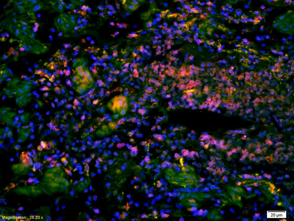
. Spindly/SPDL1 was detected in an immunocytochemical section of SiHa cells. Enzyme antigen retrieval was performed using IHC enzyme antigen retrieval reagent (AR0022) for 15 mins. The cells were blocked with 10% goat serum. And then incubated with 5 microg/mL rabbit anti-Spindly/SPDL1 Antibody (A12722-2) overnight at 4°C. Cy3 Conjugated Goat Anti-Rabbit IgG (BA1032) was used as secondary antibody at 1:500 dilution and incubated for 30 minutes at 37°C. The section was counterstained with DAPI. Visualize using a fluorescence microscope and filter sets appropriate for the label used.")
. Overlay histogram showing HCT116 cells stained with A12722-2 (Blue line). To facilitate intracellular staining, cells were fixed with 4% paraformaldehyde and permeabilized with permeabilization buffer. The cells were blocked with 10% normal goat serum. And then incubated with rabbit anti-Spindly/SPDL1 Antibody (A12722-2, 1 microg/1x106 cells) for 30 min at 20°C. DyLight®488 conjugated goat anti-rabbit IgG (BA1127, 5-10 microg/1x106 cells) was used as secondary antibody for 30 minutes at 20°C. Isotype control antibody (Green line) was rabbit IgG (1 microg/1x106) used under the same conditions. Unlabelled sample without incubation with primary antibody and secondary antibody (Red line) was used as a blank control.")
. Overlay histogram showing PC-3 cells stained with A12722-2 (Blue line). To facilitate intracellular staining, cells were fixed with 4% paraformaldehyde and permeabilized with permeabilization buffer. The cells were blocked with 10% normal goat serum. And then incubated with rabbit anti-Spindly/SPDL1 Antibody (A12722-2, 1 microg/1x106 cells) for 30 min at 20°C. DyLight®488 conjugated goat anti-rabbit IgG (BA1127, 5-10 microg/1x106 cells) was used as secondary antibody for 30 minutes at 20°C. Isotype control antibody (Green line) was rabbit IgG (1 microg/1x106) used under the same conditions. Unlabelled sample without incubation with primary antibody and secondary antibody (Red line) was used as a blank control.")
Figure 1. Western blot analysis of Spindly/SPDL1 using anti-Spindly/SPDL1 antibody (A12722-2). Electrophoresis was performed on a 5-20% SDS-PAGE gel at 70V (Stacking gel) / 90V (Resolving gel) for 2-3 hours. The sample well of each lane was loaded with 30 ug of sample under reducing conditions. Lane 1: human U251 whole cell lysates, Lane 2: human Hela whole cell lysates, Lane 3: human Caco-2 whole cell lysates, Lane 4: rat liver tissue lysates, Lane 5: rat RH35 whole cell lysates, Lane 6: mouse liver tissue lysates, Lane 7: mouse NIH/3T3 whole cell lysates. After electrophoresis, proteins were transferred to a nitrocellulose membrane at 150 mA for 50-90 minutes. Blocked the membrane with 5% non-fat milk/TBS for 1.5 hour at RT. The membrane was incubated with rabbit anti-Spindly/SPDL1 antigen affinity purified polyclonal antibody (Catalog # A12722-2) at 0.5 microg/mL overnight at 4°C, then washed with TBS-0.1%Tween 3 times with 5 minutes each and probed with a goat anti-rabbit IgG-HRP secondary antibody at a dilution of 1:5000 for 1.5 hour at RT. The signal is developed using an Enhanced Chemiluminescent detection (ECL) kit (Catalog # EK1002) with Tanon 5200 system. A specific band was detected for Spindly/SPDL1 at approximately 70 kDa. The expected band size for Spindly/SPDL1 is at 70 kDa.
Anti-Spindly/SPDL1 Antibody Picoband(r)
A12722-2-CARRIER-FREE
ApplicationsFlow Cytometry, ImmunoFluorescence, Western Blot, ELISA, ImmunoCytoChemistry
Product group Antibodies
ReactivityHuman, Mouse, Rat
TargetSPDL1
Overview
- SupplierBoster Bio
- Product NameAnti-Spindly/SPDL1 Antibody Picoband(r)
- Delivery Days Customer9
- ApplicationsFlow Cytometry, ImmunoFluorescence, Western Blot, ELISA, ImmunoCytoChemistry
- CertificationResearch Use Only
- ClonalityPolyclonal
- Concentration500 ug/ml
- Gene ID54908
- Target nameSPDL1
- Target descriptionspindle apparatus coiled-coil protein 1
- Target synonymsCCDC99, protein Spindly, arsenite-related gene 1 protein, coiled-coil domain-containing protein 99, rhabdomyosarcoma antigen MU-RMS-40.4A, rrhabdomyosarcoma antigen protein MU-RMS-40.4A
- HostRabbit
- IsotypeIgG
- Protein IDQ96EA4
- Protein NameProtein Spindly
- Scientific DescriptionBoster Bio Anti-Spindly/SPDL1 Antibody Picoband® catalog # A12722-2. Tested in ELISA, Flow Cytometry, IF, ICC, WB applications. This antibody reacts with Human, Mouse, Rat. The brand Picoband indicates this is a premium antibody that guarantees superior quality, high affinity, and strong signals with minimal background in Western blot applications. Only our best-performing antibodies are designated as Picoband, ensuring unmatched performance.
- ReactivityHuman, Mouse, Rat
- Storage Instruction-20°C,2°C to 8°C
- UNSPSC12352203
Related products
Product group Antibodies
Anti-CCDC99 (C-term) Antibody102-21493
ApplicationsWestern Blot, ImmunoHistoChemistry, ImmunoHistoChemistry Paraffin
TargetSPDL1
- SizePrice
Product group Antibodies
Anti-CCDC99 AntibodyA100598
ApplicationsWestern Blot, ELISA
ReactivityHuman
- SizePrice
Product group Antibodies
Spindly Polyclonal AntibodyBS-2321R
ApplicationsImmunoFluorescence, ELISA, ImmunoCytoChemistry, ImmunoHistoChemistry, ImmunoHistoChemistry Frozen, ImmunoHistoChemistry Paraffin
ReactivityBovine, Canine, Human, Mouse, Porcine, Rat
TargetSPDL1
- SizePrice
Product group Antibodies
SPDL1 Polyclonal AntibodyCAC14317
ApplicationsWestern Blot, ELISA, ImmunoHistoChemistry
TargetSPDL1
- SizePrice
Product group Antibodies
SPDL1 AntibodyCSB-PA030153
ApplicationsWestern Blot, ELISA
ReactivityHuman
TargetSPDL1
- SizePrice
Product group Antibodies

SPDL1 / CCDC99 AntibodyLS-C401050
ApplicationsWestern Blot, ELISA
ReactivityHuman, Mouse
TargetSPDL1
- SizePrice

Product group Antibodies
Anti-SPDL1 AntibodyHPA044700
ApplicationsImmunoHistoChemistry
ReactivityHuman
TargetSPDL1
- SizePrice
Product group Antibodies
Spindly antibodyGTX51534
ApplicationsImmunoHistoChemistry, ImmunoHistoChemistry Paraffin
ReactivityHuman
TargetSPDL1
- SizePrice