

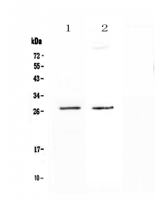
Figure 1. Western blot analysis of SPR using anti-SPR antibody (A00416-1). Electrophoresis was performed on a 5-20% SDS-PAGE gel at 70V (Stacking gel) / 90V (Resolving gel) for 2-3 hours. The sample well of each lane was loaded with 50ug of sample under reducing conditions. Lane 1: human HepG2 whole cell lysates, Lane 2: mouse HEPA1-6 whole cell lysates. After Electrophoresis, proteins were transferred to a Nitrocellulose membrane at 150mA for 50-90 minutes. Blocked the membrane with 5% Non-fat Milk/ TBS for 1.5 hour at RT. The membrane was incubated with rabbit anti-SPR antigen affinity purified polyclonal antibody (Catalog # A00416-1) at 0.5 microg/mL overnight at 4°C, then washed with TBS-0.1%Tween 3 times with 5 minutes each and probed with a goat anti-rabbit IgG-HRP secondary antibody at a dilution of 1:10000 for 1.5 hour at RT. The signal is developed using an Enhanced Chemiluminescent detection (ECL) kit (Catalog # EK1002) with Tanon 5200 system. A specific band was detected for SPR at approximately 28KD. The expected band size for SPR is at 28KD.
Figure 1. Western blot analysis of SPR using anti-SPR antibody (A00416-1). Electrophoresis was performed on a 5-20% SDS-PAGE gel at 70V (Stacking gel) / 90V (Resolving gel) for 2-3 hours. The sample well of each lane was loaded with 50ug of sample under reducing conditions. Lane 1: human HepG2 whole cell lysates, Lane 2: mouse HEPA1-6 whole cell lysates. After Electrophoresis, proteins were transferred to a Nitrocellulose membrane at 150mA for 50-90 minutes. Blocked the membrane with 5% Non-fat Milk/ TBS for 1.5 hour at RT. The membrane was incubated with rabbit anti-SPR antigen affinity purified polyclonal antibody (Catalog # A00416-1) at 0.5 microg/mL overnight at 4°C, then washed with TBS-0.1%Tween 3 times with 5 minutes each and probed with a goat anti-rabbit IgG-HRP secondary antibody at a dilution of 1:10000 for 1.5 hour at RT. The signal is developed using an Enhanced Chemiluminescent detection (ECL) kit (Catalog # EK1002) with Tanon 5200 system. A specific band was detected for SPR at approximately 28KD. The expected band size for SPR is at 28KD.
Anti-SPR Antibody Picoband(r)
A00416-1-CARRIER-FREE
ApplicationsWestern Blot, ELISA
Product group Antibodies
ReactivityHuman, Mouse
TargetSPR
Overview
- SupplierBoster Bio
- Product NameAnti-SPR Antibody Picoband(r)
- Delivery Days Customer9
- ApplicationsWestern Blot, ELISA
- CertificationResearch Use Only
- ClonalityPolyclonal
- Concentration500 ug/ml
- Gene ID6697
- Target nameSPR
- Target descriptionsepiapterin reductase
- Target synonymsSDR38C1, sepiapterin reductase, Sepiapterin reductase (L-erythro-7,8-dihydrobiopterin forming), sepiapterin reductase (7,8-dihydrobiopterin:NADP+ oxidoreductase), short chain dehydrogenase/reductase family 38C, member 1
- HostRabbit
- IsotypeIgG
- Protein IDP35270
- Protein NameSepiapterin reductase
- Scientific DescriptionBoster Bio Anti-SPR Antibody Picoband® catalog # A00416-1. Tested in ELISA, WB applications. This antibody reacts with Human, Mouse. The brand Picoband indicates this is a premium antibody that guarantees superior quality, high affinity, and strong signals with minimal background in Western blot applications. Only our best-performing antibodies are designated as Picoband, ensuring unmatched performance.
- ReactivityHuman, Mouse
- Storage Instruction-20°C,2°C to 8°C
- UNSPSC12352203
Related products
Product group Antibodies
Anti-SPR Antibody144-11694
ApplicationsWestern Blot
ReactivityHuman, Mouse, Rat
TargetSPR
- SizePrice
Product group Antibodies
ApplicationsImmunoPrecipitation, Western Blot, ImmunoCytoChemistry, ImmunoHistoChemistry
TargetSPR
- SizePrice
Product group Antibodies
SPR AntibodyCSB-PA214577
ApplicationsWestern Blot, ELISA, ImmunoHistoChemistry
ReactivityHuman, Mouse
TargetSPR
- SizePrice
Product group Antibodies

SPR AntibodyLS-C402950
ApplicationsWestern Blot, ELISA, ImmunoHistoChemistry
ReactivityHuman
TargetSPR
- SizePrice

Product group Antibodies
Anti-SPR AntibodyHPA039505
ApplicationsWestern Blot, ImmunoCytoChemistry, ImmunoHistoChemistry
ReactivityHuman
TargetSPR
- SizePrice
![SPR antibody [N2C3] immunoprecipitates SPR protein in IP experiments. IP samples: A431 whole cell extract A. Control with 4 μg of preimmune Rabbit IgG B. Immunoprecipitation of SPR protein by 4 μg SPR antibody [N2C3] (GTX113552) 5 % SDS-PAGE The immunoprecipitated SPR protein was detected by SPR antibody [N2C3] (GTX113552) diluted at 1:500. [EasyBlot anti-rabbit IgG (GTX221666-01) was used as a secondary reagent]](https://www.genetex.com/upload/website/prouct_img/normal/GTX113552/GTX113552_40142_IP_w_23060501_454.webp)
Product group Antibodies
SPR antibody [N2C3]GTX113552
ApplicationsImmunoFluorescence, ImmunoPrecipitation, Western Blot, ImmunoCytoChemistry, ImmunoHistoChemistry, ImmunoHistoChemistry Paraffin
ReactivityHuman, Mouse
TargetSPR
- SizePrice
Product group Antibodies
Anti-SPR AntibodyCAB11694
ApplicationsWestern Blot, ELISA
ReactivityHuman
TargetSPR
- SizePrice