


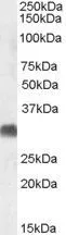
Figure 1. Western blot analysis of SRD5A2 using anti-SRD5A2 antibody (M00704). Electrophoresis was performed on a 5-20% SDS-PAGE gel at 70V (Stacking gel) / 90V (Resolving gel) for 2-3 hours. The sample well of each lane was loaded with 30 ug of sample under reducing conditions. Lane 1: human PC-3 whole cell lysates, Lane 2: human RT4 whole cell lysates, Lane 3: human SH-SY5Y whole cell lysates, Lane 4: human HepG2 whole cell lysates, Lane 5: human NIH/3T3 whole cell lysates, Lane 6: mouse HT22 whole cell lysates. After electrophoresis, proteins were transferred to a nitrocellulose membrane at 150 mA for 50-90 minutes. Blocked the membrane with 5% non-fat milk/TBS for 1.5 hour at RT. The membrane was incubated with rabbit anti-SRD5A2 antigen affinity purified monoclonal antibody (Catalog # M00704) at 1:500 overnight at 4°C, then washed with TBS-0.1%Tween 3 times with 5 minutes each and probed with a goat anti-rabbit IgG-HRP secondary antibody at a dilution of 1:500 for 1.5 hour at RT. The signal is developed using an Enhanced Chemiluminescent detection (ECL) kit (Catalog # EK1002) with Tanon 5200 system. A specific band was detected for SRD5A2 at approximately 28 kDa. The expected band size for SRD5A2 is at 28 kDa.
Figure 1. Western blot analysis of SRD5A2 using anti-SRD5A2 antibody (M00704). Electrophoresis was performed on a 5-20% SDS-PAGE gel at 70V (Stacking gel) / 90V (Resolving gel) for 2-3 hours. The sample well of each lane was loaded with 30 ug of sample under reducing conditions. Lane 1: human PC-3 whole cell lysates, Lane 2: human RT4 whole cell lysates, Lane 3: human SH-SY5Y whole cell lysates, Lane 4: human HepG2 whole cell lysates, Lane 5: human NIH/3T3 whole cell lysates, Lane 6: mouse HT22 whole cell lysates. After electrophoresis, proteins were transferred to a nitrocellulose membrane at 150 mA for 50-90 minutes. Blocked the membrane with 5% non-fat milk/TBS for 1.5 hour at RT. The membrane was incubated with rabbit anti-SRD5A2 antigen affinity purified monoclonal antibody (Catalog # M00704) at 1:500 overnight at 4°C, then washed with TBS-0.1%Tween 3 times with 5 minutes each and probed with a goat anti-rabbit IgG-HRP secondary antibody at a dilution of 1:500 for 1.5 hour at RT. The signal is developed using an Enhanced Chemiluminescent detection (ECL) kit (Catalog # EK1002) with Tanon 5200 system. A specific band was detected for SRD5A2 at approximately 28 kDa. The expected band size for SRD5A2 is at 28 kDa.
Anti-SRD5A2 Rabbit Monoclonal Antibody
M00704
ApplicationsImmunoPrecipitation, Western Blot
Product group Antibodies
ReactivityHuman, Mouse, Rat
TargetSRD5A2
Overview
- SupplierBoster Bio
- Product NameAnti-SRD5A2 Rabbit Monoclonal Antibody
- Delivery Days Customer9
- ApplicationsImmunoPrecipitation, Western Blot
- CertificationResearch Use Only
- ClonalityMonoclonal
- Clone ID24S47
- Gene ID6716
- Target nameSRD5A2
- Target descriptionsteroid 5 alpha-reductase 2
- Target synonyms3-oxo-5-alpha-steroid 4-dehydrogenase 2, 5 alpha-SR2, S5AR 2, SR type 2, steroid-5-alpha-reductase, alpha polypeptide 2 (3-oxo-5 alpha-steroid delta 4-dehydrogenase alpha 2), type II 5-alpha reductase
- HostRabbit
- IsotypeIgG
- Protein IDP31213
- Protein Name3-oxo-5-alpha-steroid 4-dehydrogenase 2
- Scientific DescriptionBoster Bio Anti-SRD5A2 Rabbit Monoclonal Antibody catalog # M00704. Tested in WB, IP applications. This antibody reacts with Human, Mouse, Rat.
- ReactivityHuman, Mouse, Rat
- Storage Instruction-20°C
- UNSPSC12352203
Related products
Product group Antibodies

SRD5A2 AntibodyLS-C780518
ApplicationsWestern Blot, ELISA
ReactivityHuman, Mouse, Rat
TargetSRD5A2
- SizePrice
Product group Antibodies

ApplicationsWestern Blot, ELISA
ReactivityHuman
TargetSRD5A2
- SizePrice
Product group Antibodies
References
SRD5A2 antibody, C-termGTX47547
ApplicationsWestern Blot
ReactivityHuman, Porcine
TargetSRD5A2
- SizePrice
Product group Antibodies
Anti-SRD5A2 (Center) Antibody102-26862
ApplicationsWestern Blot
TargetSRD5A2
- SizePrice
Product group Antibodies
References
SRD5A2 Polyclonal AntibodyBS-6700R
ApplicationsImmunoFluorescence, ELISA, ImmunoHistoChemistry, ImmunoHistoChemistry Frozen, ImmunoHistoChemistry Paraffin
ReactivityHuman, Mouse, Rat
TargetSRD5A2
- SizePrice