Bio-Connect
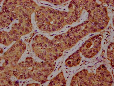
Immunohistochemical staining of human kidney shows strong cytoplasmic positivity in cells in tubules.

Anti-SSH1 Antibody

HPA019845
Atlas Antibodies
ApplicationsImmunoHistoChemistry
Product group Antibodies
TargetSSH1
Sign in to order and to see your custom pricing.
Large volume orders?
Order with a bulk request

Overview

  • Supplier
    Atlas Antibodies
  • Product Name
    Anti-SSH1
  • Delivery Days Customer
    4
  • Applications
    ImmunoHistoChemistry
  • Certification
    Research Use Only
  • Clonality
    Polyclonal
  • Conjugate
    Unconjugated
  • Gene ID54434
  • Target name
    SSH1
  • Target description
    slingshot protein phosphatase 1
  • Target synonyms
    hSSH-1L; protein phosphatase Slingshot homolog 1; slingshot homolog 1; SSH1L; SSH-like protein 1
  • Host
    Rabbit
  • Isotype
    IgG
  • Protein IDQ8WYL5
  • Protein Name
    Protein phosphatase Slingshot homolog 1
  • Scientific Description
    Recombinant Protein Epitope Signature Tag (PrEST) antigen sequence
  • Storage Instruction
    -20°C,2°C to 8°C
  • UNSPSC
    12352203

Related products

IHC image of CSB-PA837881LA01HU diluted at 1:300 and staining in paraffin-embedded human liver cancer performed on a Leica BondTM system. After dewaxing and hydration, antigen retrieval was mediated by high pressure in a citrate buffer (pH 6.0). Section was blocked with 10% normal goat serum 30min at RT. Then primary antibody (1% BSA) was incubated at 4°C overnight. The primary is detected by a biotinylated secondary antibody and visualized using an HRP conjugated SP system.
Product group Antibodies
Cusabio
SSH1 AntibodyCSB-PA837881LA01HU
ApplicationsImmunoFluorescence, ELISA, ImmunoHistoChemistry
TargetSSH1
  • SizePrice
Product group Antibodies
RayBiotech
ApplicationsWestern Blot
TargetSSH1
  • SizePrice
Atlas Antibodies
  • SizePrice
Product group Antibodies
Bethyl Laboratories
ApplicationsImmunoPrecipitation, Western Blot
TargetSSH1
  • SizePrice
Figure 1. Western blot analysis of SSH1 using anti-SSH1 antibody (A03480). Electrophoresis was performed on a 5-20% SDS-PAGE gel at 70V (Stacking gel) / 90V (Resolving gel) for 2-3 hours. The sample well of each lane was loaded with 30 ug of sample under reducing conditions. Lane 1: human Hela whole cell lysates, Lane 2: human 293T whole cell lysates, Lane 3: human Jurkat whole cell lysates, Lane 4: human Raji whole cell lysates. After electrophoresis, proteins were transferred to a nitrocellulose membrane at 150 mA for 50-90 minutes. Blocked the membrane with 5% non-fat milk/TBS for 1.5 hour at RT. The membrane was incubated with rabbit anti-SSH1 antigen affinity purified polyclonal antibody (Catalog # A03480) at 0.5 microg/mL overnight at 4°C, then washed with TBS-0.1%Tween 3 times with 5 minutes each and probed with a goat anti-rabbit IgG-HRP secondary antibody at a dilution of 1:5000 for 1.5 hour at RT. The signal is developed using an Enhanced Chemiluminescent detection (ECL) kit (Catalog # EK1002) with Tanon 5200 system. A specific band was detected for SSH1 at approximately 150 kDa. The expected band size for SSH1 is at 116 kDa.
Product group Antibodies
Boster Bio
ApplicationsFlow Cytometry, ImmunoFluorescence, Western Blot, ELISA, ImmunoCytoChemistry
TargetSSH1
  • SizePrice
Product group Antibodies
LifeSpan BioSciences
SSH1 AntibodyLS-C681465
ApplicationsImmunoFluorescence, ELISA, ImmunoHistoChemistry, ImmunoHistoChemistry Paraffin
TargetSSH1
  • SizePrice
Product group Antibodies
Assay Genie
SSH1 AntibodyPACO60248
ApplicationsImmunoFluorescence, ELISA, ImmunoHistoChemistry
TargetSSH1
  • SizePrice
Product group Antibodies
Bioss Antibodies
ApplicationsWestern Blot, ImmunoHistoChemistry, ImmunoHistoChemistry Paraffin
TargetSSH1
  • SizePrice