


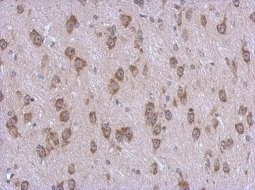
Immunohistochemical staining of human stomach shows strong cytoplasmic and membranous positivity in glandular cells.

Immunohistochemical staining of human stomach shows strong cytoplasmic and membranous positivity in glandular cells.
Anti-STAMBPL1 Antibody
HPA040202
ApplicationsImmunoCytoChemistry, ImmunoHistoChemistry
Product group Antibodies
ReactivityHuman
TargetSTAMBPL1
Overview
- SupplierAtlas Antibodies
- Product NameAnti-STAMBPL1 Antibody
- Delivery Days Customer4
- ApplicationsImmunoCytoChemistry, ImmunoHistoChemistry
- CertificationResearch Use Only
- ClonalityPolyclonal
- ConjugateUnconjugated
- Gene ID57559
- Target nameSTAMBPL1
- Target descriptionSTAM binding protein like 1
- Target synonymsALMalpha, AMSH-FP, AMSH-LP, bA399O19.2, AMSH-like protease, associated molecule with the SH3 domain of STAM (AMSH) - Family Protein, associated molecule with the SH3 domain of STAM (AMSH) like protein
- HostRabbit
- IsotypeIgG
- Protein IDQ96FJ0
- Protein NameAMSH-like protease
- Scientific DescriptionRecombinant Protein Epitope Signature Tag (PrEST) antigen sequence
- ReactivityHuman
- Storage Instruction-20°C,2°C to 8°C
- UNSPSC41116161
Datasheet
MSDS
Related products

Product group Antibodies
Anti-AMSH-LP/STAMBPL1 Antibody Picoband(r)A09862-1-CARRIER-FREE
ApplicationsFlow Cytometry, Western Blot, ELISA
ReactivityHuman, Mouse
TargetSTAMBPL1
- SizePrice
Product group Antibodies
Anti-STAMBPL1 Antibody144-63955
ApplicationsWestern Blot
ReactivityHuman, Mouse, Rat
TargetSTAMBPL1
- SizePrice
Product group Antibodies
STAMBPL1 antibody [N3C3]GTX120491
ApplicationsImmunoFluorescence, Western Blot, ImmunoCytoChemistry, ImmunoHistoChemistry, ImmunoHistoChemistry Paraffin
ReactivityHuman
TargetSTAMBPL1
- SizePrice
Product group Antibodies

STAMBPL1 Antibody (aa135-163)LS-C161540
ApplicationsFlow Cytometry, Western Blot, ImmunoHistoChemistry, ImmunoHistoChemistry Paraffin
ReactivityHuman, Mouse
TargetSTAMBPL1
- SizePrice