


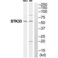
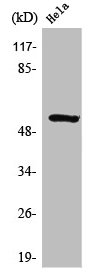
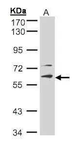
Figure 1. Western blot analysis of STK33 using anti-STK33 antibody (M07375). Electrophoresis was performed on a 5-20% SDS-PAGE gel at 70V (Stacking gel) / 90V (Resolving gel) for 2-3 hours. The sample well of each lane was loaded with 30 ug of sample under reducing conditions. Lane 1: human 293T whole cell lysates, Lane 2: human U251 whole cell lysates, Lane 3: human Jurkat whole cell lysates, Lane 4: human U2OS whole cell lysates, Lane 5: human Hela whole cell lysates, Lane 6: human HepG2 whole cell lysates, Lane 7: human A549 whole cell lysates. After electrophoresis, proteins were transferred to a nitrocellulose membrane at 150 mA for 50-90 minutes. Blocked the membrane with 5% non-fat milk/TBS for 1.5 hour at RT. The membrane was incubated with rabbit anti-STK33 antigen affinity purified monoclonal antibody (M07375) at 1:500 overnight at 4°C, then washed with TBS-0.1%Tween 3 times with 5 minutes each and probed with a goat anti-rabbit IgG-HRP secondary antibody at a dilution of 1:500 for 1.5 hour at RT. The signal is developed using an Enhanced Chemiluminescent detection (ECL) kit (Catalog # EK1002) with Tanon 5200 system. A specific band was detected for STK33 at approximately 58 kDa. The expected band size for STK33 is at 58, 50 kDa.
Figure 1. Western blot analysis of STK33 using anti-STK33 antibody (M07375). Electrophoresis was performed on a 5-20% SDS-PAGE gel at 70V (Stacking gel) / 90V (Resolving gel) for 2-3 hours. The sample well of each lane was loaded with 30 ug of sample under reducing conditions. Lane 1: human 293T whole cell lysates, Lane 2: human U251 whole cell lysates, Lane 3: human Jurkat whole cell lysates, Lane 4: human U2OS whole cell lysates, Lane 5: human Hela whole cell lysates, Lane 6: human HepG2 whole cell lysates, Lane 7: human A549 whole cell lysates. After electrophoresis, proteins were transferred to a nitrocellulose membrane at 150 mA for 50-90 minutes. Blocked the membrane with 5% non-fat milk/TBS for 1.5 hour at RT. The membrane was incubated with rabbit anti-STK33 antigen affinity purified monoclonal antibody (M07375) at 1:500 overnight at 4°C, then washed with TBS-0.1%Tween 3 times with 5 minutes each and probed with a goat anti-rabbit IgG-HRP secondary antibody at a dilution of 1:500 for 1.5 hour at RT. The signal is developed using an Enhanced Chemiluminescent detection (ECL) kit (Catalog # EK1002) with Tanon 5200 system. A specific band was detected for STK33 at approximately 58 kDa. The expected band size for STK33 is at 58, 50 kDa.
Anti-STK33 Rabbit Monoclonal Antibody
M07375
ApplicationsImmunoFluorescence, Western Blot, ImmunoCytoChemistry, ImmunoHistoChemistry
Product group Antibodies
ReactivityHuman
TargetSTK33
Overview
- SupplierBoster Bio
- Product NameAnti-STK33 Rabbit Monoclonal Antibody
- Delivery Days Customer9
- ApplicationsImmunoFluorescence, Western Blot, ImmunoCytoChemistry, ImmunoHistoChemistry
- CertificationResearch Use Only
- ClonalityMonoclonal
- Clone ID22S21
- Gene ID65975
- Target nameSTK33
- Target descriptionserine/threonine kinase 33
- Target synonymsSPGF93, serine/threonine-protein kinase 33
- HostRabbit
- IsotypeIgG
- Protein IDQ9BYT3
- Protein NameSerine/threonine-protein kinase 33
- Scientific DescriptionBoster Bio Anti-STK33 Rabbit Monoclonal Antibody catalog # M07375. Tested in WB, IHC, ICC/IF applications. This antibody reacts with Human.
- ReactivityHuman
- Storage Instruction-20°C
- UNSPSC12352203
Related products
Product group Antibodies
STK33 Recombinant Antibody, AbBy Fluor-405 ConjugatedBSM-62085R-BF405
ApplicationsImmunoFluorescence, Western Blot
ReactivityHuman
TargetSTK33
- SizePrice
Product group Antibodies
STK33 AntibodyCSB-PA004188
ApplicationsWestern Blot, ELISA, ImmunoHistoChemistry
ReactivityHuman
TargetSTK33
- SizePrice
Product group Antibodies
STK33 Polyclonal AntibodyCAC14387
ApplicationsImmunoFluorescence, Western Blot, ELISA, ImmunoHistoChemistry
ReactivityMouse
TargetSTK33
- SizePrice
Product group Antibodies

STK33 AntibodyLS-C379296
ApplicationsWestern Blot, ELISA, ImmunoHistoChemistry, ImmunoHistoChemistry Paraffin
ReactivityHuman, Mouse
TargetSTK33
- SizePrice
Product group Antibodies
STK33 antibody [N1C1]GTX105293
ApplicationsWestern Blot
ReactivityHuman
TargetSTK33
- SizePrice

Product group Antibodies
Anti-STK33 AntibodyHPA015742
ApplicationsImmunoHistoChemistry
ReactivityHuman
TargetSTK33
- SizePrice