


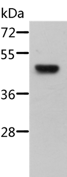
Figure 1. Western blot analysis of STRADB using anti-STRADB antibody (A05271-2). Electrophoresis was performed on a 5-20% SDS-PAGE gel at 70V (Stacking gel) / 90V (Resolving gel) for 2-3 hours. The sample well of each lane was loaded with 30 ug of sample under reducing conditions. Lane 1: human Raji whole cell lysates, Lane 2: human HepG2 whole cell lysates, Lane 3: human SH-SY5Y whole cell lysates, Lane 4: human K562 whole cell lysates. After electrophoresis, proteins were transferred to a nitrocellulose membrane at 150 mA for 50-90 minutes. Blocked the membrane with 5% non-fat milk/TBS for 1.5 hour at RT. The membrane was incubated with rabbit anti-STRADB antigen affinity purified polyclonal antibody (Catalog # A05271-2) at 0.5 microg/mL overnight at 4°C, then washed with TBS-0.1%Tween 3 times with 5 minutes each and probed with a goat anti-rabbit IgG-HRP secondary antibody at a dilution of 1:5000 for 1.5 hour at RT. The signal is developed using an Enhanced Chemiluminescent detection (ECL) kit (Catalog # EK1002) with Tanon 5200 system. A specific band was detected for STRADB at approximately 33 kDa. The expected band size for STRADB is at 47 kDa.
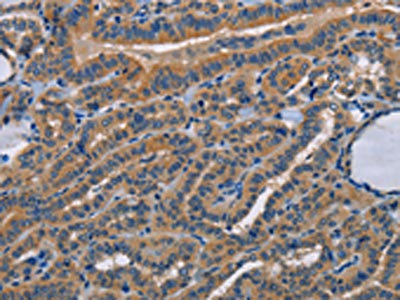
. STRADB was detected in an immunocytochemical section of A549 cells. Enzyme antigen retrieval was performed using IHC enzyme antigen retrieval reagent (AR0022) for 15 mins. The cells were blocked with 10% goat serum. And then incubated with 5 microg/mL rabbit anti-STRADB Antibody (A05271-2) overnight at 4°C. Cy3 Conjugated Goat Anti-Rabbit IgG (BA1032) was used as secondary antibody at 1:500 dilution and incubated for 30 minutes at 37°C. The section was counterstained with DAPI. Visualize using a fluorescence microscope and filter sets appropriate for the label used.")
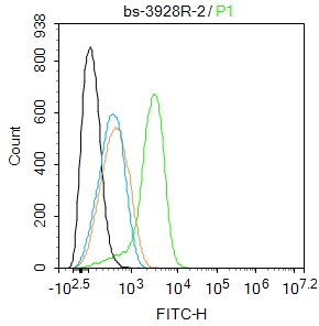
. Overlay histogram showing SiHa cells stained with A05271-2 (Blue line). To facilitate intracellular staining, cells were fixed with 4% paraformaldehyde and permeabilized with permeabilization buffer. The cells were blocked with 10% normal goat serum. And then incubated with rabbit anti-STRADB Antibody (A05271-2, 1 microg/1x106 cells) for 30 min at 20°C. DyLight®488 conjugated goat anti-rabbit IgG (BA1127, 5-10 microg/1x106 cells) was used as secondary antibody for 30 minutes at 20°C. Isotype control antibody (Green line) was rabbit IgG (1 microg/1x106) used under the same conditions. Unlabelled sample (Red line) was also used as a control.")
Figure 1. Western blot analysis of STRADB using anti-STRADB antibody (A05271-2). Electrophoresis was performed on a 5-20% SDS-PAGE gel at 70V (Stacking gel) / 90V (Resolving gel) for 2-3 hours. The sample well of each lane was loaded with 30 ug of sample under reducing conditions. Lane 1: human Raji whole cell lysates, Lane 2: human HepG2 whole cell lysates, Lane 3: human SH-SY5Y whole cell lysates, Lane 4: human K562 whole cell lysates. After electrophoresis, proteins were transferred to a nitrocellulose membrane at 150 mA for 50-90 minutes. Blocked the membrane with 5% non-fat milk/TBS for 1.5 hour at RT. The membrane was incubated with rabbit anti-STRADB antigen affinity purified polyclonal antibody (Catalog # A05271-2) at 0.5 microg/mL overnight at 4°C, then washed with TBS-0.1%Tween 3 times with 5 minutes each and probed with a goat anti-rabbit IgG-HRP secondary antibody at a dilution of 1:5000 for 1.5 hour at RT. The signal is developed using an Enhanced Chemiluminescent detection (ECL) kit (Catalog # EK1002) with Tanon 5200 system. A specific band was detected for STRADB at approximately 33 kDa. The expected band size for STRADB is at 47 kDa.
Anti-STRADB Antibody Picoband(r)
A05271-2-CARRIER-FREE
ApplicationsFlow Cytometry, ImmunoFluorescence, Western Blot, ELISA, ImmunoCytoChemistry
Product group Antibodies
ReactivityHuman
TargetSTRADB
Overview
- SupplierBoster Bio
- Product NameAnti-STRADB Antibody Picoband(r)
- Delivery Days Customer9
- ApplicationsFlow Cytometry, ImmunoFluorescence, Western Blot, ELISA, ImmunoCytoChemistry
- CertificationResearch Use Only
- ClonalityPolyclonal
- Concentration500 ug/ml
- Gene ID55437
- Target nameSTRADB
- Target descriptionSTE20 related adaptor beta
- Target synonymsALS2CR2, CALS-21, ILPIP, ILPIPA, PAPK, PRO1038, STE20-related kinase adapter protein beta, ILP-interacting protein ILPIPA, STE20-related kinase adaptor beta, STRAD beta, amyotrophic lateral sclerosis 2 (juvenile) chromosome region, candidate 2, amyotrophic lateral sclerosis 2 chromosomal region candidate gene 2 protein, pseudokinase ALS2CR2
- HostRabbit
- IsotypeIgG
- Protein IDQ9C0K7
- Protein NameSTE20-related kinase adapter protein beta
- Scientific DescriptionBoster Bio Anti-STRADB Antibody Picoband® catalog # A05271-2. Tested in ELISA, Flow Cytometry, IF, ICC, WB applications. This antibody reacts with Human. The brand Picoband indicates this is a premium antibody that guarantees superior quality, high affinity, and strong signals with minimal background in Western blot applications. Only our best-performing antibodies are designated as Picoband, ensuring unmatched performance.
- ReactivityHuman
- Storage Instruction-20°C,2°C to 8°C
- UNSPSC12352203
Related products
Product group Antibodies
Anti-STRADB AntibodyA37191
ApplicationsWestern Blot, ImmunoHistoChemistry
ReactivityHuman, Mouse
- SizePrice
Product group Antibodies
Anti-ALS2CR2 (C-term L289) Antibody102-27175
ApplicationsWestern Blot
TargetSTRADB
- SizePrice
Product group Antibodies
ALS2CR2 Polyclonal AntibodyBS-3928R
ApplicationsImmunoFluorescence, Western Blot, ELISA, ImmunoCytoChemistry, ImmunoHistoChemistry, ImmunoHistoChemistry Frozen, ImmunoHistoChemistry Paraffin
ReactivityHuman, Mouse, Rat
TargetSTRADB
- SizePrice
Product group Antibodies

Goat anti-ALS2CR2 / ILPIPEB05865
ApplicationsImmunoFluorescence, Western Blot, ELISA, ImmunoHistoChemistry
ReactivityBovine, Canine, Human
TargetSTRADB
- SizePrice
Product group Antibodies
STRADB Polyclonal AntibodyCAC14432
ApplicationsWestern Blot, ELISA, ImmunoHistoChemistry
TargetSTRADB
- SizePrice
Product group Antibodies
STRADB AntibodyCSB-PA052910
ApplicationsWestern Blot, ELISA, ImmunoHistoChemistry
ReactivityHuman, Mouse
TargetSTRADB
- SizePrice
Product group Antibodies

STRADB / ALS2CR2 AntibodyLS-C401232
ApplicationsWestern Blot, ELISA, ImmunoHistoChemistry
ReactivityHuman, Mouse
TargetSTRADB
- SizePrice

Product group Antibodies
Anti-STRADB AntibodyHPA026549
ApplicationsWestern Blot, ImmunoCytoChemistry, ImmunoHistoChemistry
ReactivityHuman, Mouse
TargetSTRADB
- SizePrice
Product group Antibodies
ALS2CR2 antibody [C3], C-termGTX107704
ApplicationsImmunoFluorescence, Western Blot, ImmunoCytoChemistry, ImmunoHistoChemistry, ImmunoHistoChemistry Paraffin
ReactivityHuman
TargetSTRADB
- SizePrice