

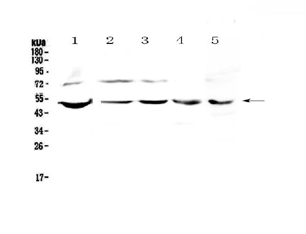
Figure 1. Western blot analysis of ESRRG using anti-ESRRG antibody (A01470-1). Electrophoresis was performed on a 5-20% SDS-PAGE gel at 70V (Stacking gel) / 90V (Resolving gel) for 2-3 hours. The sample well of each lane was loaded with 50ug of sample under reducing conditions. Lane 1: human PANC-1 whole cell lysates, Lane 2: human 22RV1 whole cell lysates, Lane 3: human COLO-320 whole cell lysates, Lane 4: human SW620 whole cell lysates, Lane 5: mouse SP20 whole cell lysates. After Electrophoresis, proteins were transferred to a Nitrocellulose membrane at 150mA for 50-90 minutes. Blocked the membrane with 5% Non-fat Milk/ TBS for 1.5 hour at RT. The membrane was incubated with rabbit anti-ESRRG antigen affinity purified polyclonal antibody (Catalog # A01470-1) at 0.5 microg/mL overnight at 4°C, then washed with TBS-0.1%Tween 3 times with 5 minutes each and probed with a goat anti-rabbit IgG-HRP secondary antibody at a dilution of 1:10000 for 1.5 hour at RT. The signal is developed using an Enhanced Chemiluminescent detection (ECL) kit (Catalog # EK1002) with Tanon 5200 system. A specific band was detected for ESRRG at approximately 51KD. The expected band size for ESRRG is at 51KD.
. ESRRG was detected in paraffin-embedded section of mouse brain tissue . Heat mediated antigen retrieval was performed in citrate buffer (pH6, epitope retrieval solution) for 20 mins. The tissue section was blocked with 10% goat serum. The tissue section was then incubated with 1microg/ml rabbit anti-ESRRG Antibody (A01470-1) overnight at 4°C. Biotinylated goat anti-rabbit IgG was used as secondary antibody and incubated for 30 minutes at 37°C. The tissue section was developed using Strepavidin-Biotin-Complex (SABC)(Catalog # SA1022) with DAB as the chromogen.")
. ESRRG was detected in paraffin-embedded section of rat cardiac muscle tissue. Heat mediated antigen retrieval was performed in citrate buffer (pH6, epitope retrieval solution) for 20 mins. The tissue section was blocked with 10% goat serum. The tissue section was then incubated with 1microg/ml rabbit anti-ESRRG Antibody (A01470-1) overnight at 4°C. Biotinylated goat anti-rabbit IgG was used as secondary antibody and incubated for 30 minutes at 37°C. The tissue section was developed using Strepavidin-Biotin-Complex (SABC)(Catalog # SA1022) with DAB as the chromogen.")
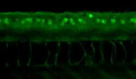
. ESRRG was detected in immunocytochemical section of A549 cell. Enzyme antigen retrieval was performed using IHC enzyme antigen retrieval reagent (AR0022) for 15 mins. The cells were blocked with 10% goat serum. And then incubated with 2microg/mL rabbit anti-ESRRG Antibody (A01470-1) overnight at 4°C. DyLight®488 Conjugated Goat Anti-Rabbit IgG (BA1127) was used as secondary antibody at 1:100 dilution and incubated for 30 minutes at 37°C. The section was counterstained with DAPI. Visualize using a fluorescence microscope and filter sets appropriate for the label used.")
. Overlay histogram showing U20S cells stained with A01470-1 (Blue line). To facilitate intracellular staining, cells were fixed with 4% paraformaldehyde and permeabilized with permeabilization buffer. The cells were blocked with 10% normal goat serum. And then incubated with rabbit anti-ESRRG Antibody (A01470-1,1microg/1x106 cells) for 30 min at 20°C. DyLight®488 conjugated goat anti-rabbit IgG (BA1127, 5-10microg/1x106 cells) was used as secondary antibody for 30 minutes at 20°C. Isotype control antibody (Green line) was rabbit IgG (1microg/1x106) used under the same conditions. Unlabelled sample without incubation with primary antibody and secondary antibody (Red line) was used as a blank control.")
Figure 1. Western blot analysis of ESRRG using anti-ESRRG antibody (A01470-1). Electrophoresis was performed on a 5-20% SDS-PAGE gel at 70V (Stacking gel) / 90V (Resolving gel) for 2-3 hours. The sample well of each lane was loaded with 50ug of sample under reducing conditions. Lane 1: human PANC-1 whole cell lysates, Lane 2: human 22RV1 whole cell lysates, Lane 3: human COLO-320 whole cell lysates, Lane 4: human SW620 whole cell lysates, Lane 5: mouse SP20 whole cell lysates. After Electrophoresis, proteins were transferred to a Nitrocellulose membrane at 150mA for 50-90 minutes. Blocked the membrane with 5% Non-fat Milk/ TBS for 1.5 hour at RT. The membrane was incubated with rabbit anti-ESRRG antigen affinity purified polyclonal antibody (Catalog # A01470-1) at 0.5 microg/mL overnight at 4°C, then washed with TBS-0.1%Tween 3 times with 5 minutes each and probed with a goat anti-rabbit IgG-HRP secondary antibody at a dilution of 1:10000 for 1.5 hour at RT. The signal is developed using an Enhanced Chemiluminescent detection (ECL) kit (Catalog # EK1002) with Tanon 5200 system. A specific band was detected for ESRRG at approximately 51KD. The expected band size for ESRRG is at 51KD.
Anti-strogen Related Receptor gamma/ESRRG Antibody Picoband(r)
A01470-1-DYLIGHT488
ApplicationsFlow Cytometry, ImmunoFluorescence, Western Blot, ELISA, ImmunoCytoChemistry, ImmunoHistoChemistry, ImmunoHistoChemistry Frozen
Product group Antibodies
ReactivityHuman, Mouse, Rat
TargetESRRG
Overview
- SupplierBoster Bio
- Product NameAnti-strogen Related Receptor gamma/ESRRG Antibody Picoband(r)
- Delivery Days Customer9
- ApplicationsFlow Cytometry, ImmunoFluorescence, Western Blot, ELISA, ImmunoCytoChemistry, ImmunoHistoChemistry, ImmunoHistoChemistry Frozen
- CertificationResearch Use Only
- ClonalityPolyclonal
- Concentration500 ug/ml
- ConjugateDyLight 488
- Gene ID2104
- Target nameESRRG
- Target descriptionestrogen related receptor gamma
- Target synonymsERR-gamma, ERR3, ERRg, ERRgamma, NR3B3, estrogen-related receptor gamma, ERR gamma-2, estrogen receptor-related protein 3, nuclear receptor subfamily 3 group B member 3
- HostRabbit
- IsotypeIgG
- Protein IDP62508
- Protein NameEstrogen-related receptor gamma
- Scientific DescriptionBoster Bio Anti-strogen Related Receptor gamma/ESRRG Antibody Picoband® catalog # A01470-1. Tested in ELISA, Flow Cytometry, IF, IHC, IHC-F, ICC, WB applications. This antibody reacts with Human, Mouse, Rat. The brand Picoband indicates this is a premium antibody that guarantees superior quality, high affinity, and strong signals with minimal background in Western blot applications. Only our best-performing antibodies are designated as Picoband, ensuring unmatched performance.
- ReactivityHuman, Mouse, Rat
- Storage Instruction-20°C,2°C to 8°C
- UNSPSC12352203
Related products
Product group Antibodies
Anti-ESRRG Antibody144-65745
ApplicationsWestern Blot
ReactivityHuman, Mouse, Rat
TargetESRRG
- SizePrice
Product group Antibodies
ApplicationsWestern Blot
ReactivityHuman, Mouse, Rat
TargetESRRG
- SizePrice
Product group Antibodies
ESRRG AntibodyCSB-PA007837LA01HU
ApplicationsELISA
ReactivityHuman
TargetESRRG
- SizePrice

Product group Antibodies
Anti-ESRRG AntibodyA37923
ApplicationsWestern Blot, ImmunoHistoChemistry
ReactivityHuman, Mouse
- SizePrice

Product group Antibodies
Anti-ESRRG AntibodyHPA044678
ApplicationsImmunoCytoChemistry, ImmunoHistoChemistry
ReactivityHuman
TargetESRRG
- SizePrice
Product group Antibodies

Goat anti-ESRRG AntibodyEB08367
ApplicationsWestern Blot, ELISA
ReactivityCanine, Human, Mouse
TargetESRRG
- SizePrice
Product group Antibodies
TargetESRRG
- SizePrice
Product group Antibodies

ESRRG / ERR Gamma AntibodyLS-C404942
ApplicationsWestern Blot, ELISA, ImmunoHistoChemistry
ReactivityHuman, Mouse, Rat
TargetESRRG
- SizePrice
Product group Antibodies
References
ERR gamma antibodyGTX102664
ApplicationsWestern Blot, ImmunoHistoChemistry
ReactivityHuman, Mouse, Rat, Zebra Fish
TargetESRRG
- SizePrice