


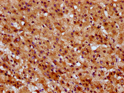
Immunohistochemical staining of human cerebellum, fallopian tube, kidney and stomach using Anti-SUCLG1 antibody HPA036684 (A) shows similar protein distribution across tissues to independent antibody HPA036683 (B).
 shows similar pattern to independent antibody HPA036683 (B).")
Immunohistochemical staining of human cerebellum, fallopian tube, kidney and stomach using Anti-SUCLG1 antibody HPA036684 (A) shows similar protein distribution across tissues to independent antibody HPA036683 (B).
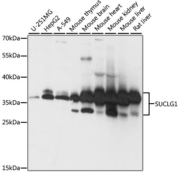
Anti-SUCLG1 Antibody
HPA036684
ApplicationsWestern Blot, ImmunoHistoChemistry
Product group Antibodies
ReactivityHuman
TargetSUCLG1
Overview
- SupplierAtlas Antibodies
- Product NameAnti-SUCLG1 Antibody
- Delivery Days Customer4
- ApplicationsWestern Blot, ImmunoHistoChemistry
- CertificationResearch Use Only
- ClonalityPolyclonal
- ConjugateUnconjugated
- Gene ID8802
- Target nameSUCLG1
- Target descriptionsuccinate-CoA ligase GDP/ADP-forming subunit alpha
- Target synonymsGALPHA, MTDPS9, SUCLA1, succinate--CoA ligase [ADP/GDP-forming] subunit alpha, mitochondrial, SCS-alpha, mitochondrial succinate--CoA ligase [ADP/GDP-forming] subunit alpha, succinate-CoA ligase alpha subunit, succinyl-CoA ligase [ADP/GDP-forming] subunit alpha, mitochondrial, succinyl-CoA ligase [GDP-forming] subunit alpha, mitochondrial, succinyl-CoA synthetase subunit alpha
- HostRabbit
- IsotypeIgG
- Protein IDP53597
- Protein NameSuccinate--CoA ligase [ADP/GDP-forming] subunit alpha, mitochondrial
- Scientific DescriptionRecombinant Protein Epitope Signature Tag (PrEST) antigen sequence
- ReactivityHuman
- Storage Instruction-20°C,2°C to 8°C
- UNSPSC41116161
Datasheet
MSDS
Related products
Product group Antibodies
Anti-SUCLG1 Antibody144-61004
ApplicationsWestern Blot
ReactivityHuman, Mouse, Rat
TargetSUCLG1
- SizePrice
Product group Antibodies
References
SUCLG1 antibodyGTX109215
ApplicationsImmunoFluorescence, ImmunoPrecipitation, Western Blot, ImmunoCytoChemistry, ImmunoHistoChemistry, ImmunoHistoChemistry Paraffin
ReactivityHuman, Mouse, Rat
TargetSUCLG1
- SizePrice
Product group Antibodies
Anti-SUCLG1 AntibodyA87637
ApplicationsImmunoFluorescence, Western Blot, ImmunoCytoChemistry
ReactivityHuman, Mouse, Rat
- SizePrice

Product group Antibodies
Anti-SUCLG1 Antibody Picoband(r)A06274-1-CARRIER-FREE
ApplicationsFlow Cytometry, ImmunoFluorescence, Western Blot, ELISA, ImmunoCytoChemistry, ImmunoHistoChemistry
ReactivityHuman, Rat
TargetSUCLG1
- SizePrice
Product group Antibodies
SUCLG1 AntibodyCSB-PA022919LA01HU
ApplicationsELISA, ImmunoHistoChemistry
ReactivityHuman
TargetSUCLG1
- SizePrice
Product group Antibodies

SUCLG1 / GALPHA AntibodyLS-C750316
ApplicationsWestern Blot
ReactivityHuman, Mouse, Rat
TargetSUCLG1
- SizePrice

Product group Antibodies
Anti-SUCLG1 AntibodyHPA036683
ApplicationsWestern Blot, ImmunoCytoChemistry, ImmunoHistoChemistry
ReactivityHuman, Mouse, Rat
TargetSUCLG1
- SizePrice