


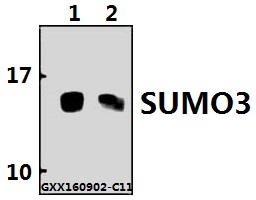
Figure 1. Western blot analysis of SUMO2/3 using anti-SUMO2/3 antibody (A01282-2). Electrophoresis was performed on a 5-20% SDS-PAGE gel at 70V (Stacking gel) / 90V (Resolving gel) for 2-3 hours. The sample well of each lane was loaded with 30 ug of sample under reducing conditions. Lane 1: human Jurkat whole cell lysates, Lane 2: human HL-60 whole cell lysates, Lane 3: human THP-1 whole cell lysates, Lane 4: human K562 whole cell lysates, Lane 5: human Caco-2 whole cell lysates, Lane 6: rat C6 whole cell lysates, Lane 8: mouse SP2/0 whole cell lysates, Lane 7: mouse NIH/3T3 whole cell lysates. After electrophoresis, proteins were transferred to a nitrocellulose membrane at 150 mA for 50-90 minutes. Blocked the membrane with 5% non-fat milk/TBS for 1.5 hour at RT. The membrane was incubated with rabbit anti-A01282-2 antigen affinity purified polyclonal antibody (Catalog # A01282-2) at 0.5 microg/mL overnight at 4°C, then washed with TBS-0.1%Tween 3 times with 5 minutes each and probed with a goat anti-rabbit IgG-HRP secondary antibody at a dilution of 1:5000 for 1.5 hour at RT. The signal is developed using an Enhanced Chemiluminescent detection (ECL) kit (Catalog # EK1002) with Tanon 5200 system. A specific band was detected for SUMO2/3 at approximately 20 kDa. The expected band size for SUMO2/3 is at 20 kDa.
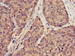
. SUMO2/3 was detected in a paraffin-embedded section of human bladder cancer tissue. Heat mediated antigen retrieval was performed in EDTA buffer (pH 8.0, epitope retrieval solution). The tissue section was blocked with 10% goat serum. The tissue section was then incubated with 2 microg/ml rabbit anti-SUMO2/3 Antibody (A01282-2) overnight at 4°C. Biotinylated goat anti-rabbit IgG was used as secondary antibody and incubated for 30 minutes at 37°C. The tissue section was developed using Strepavidin-Biotin-Complex (SABC) (Catalog # SA1022) with DAB as the chromogen.")
. SUMO2/3 was detected in a paraffin-embedded section of human gallbladder adenocarcinoma tissue. Heat mediated antigen retrieval was performed in EDTA buffer (pH 8.0, epitope retrieval solution). The tissue section was blocked with 10% goat serum. The tissue section was then incubated with 2 microg/ml rabbit anti-SUMO2/3 Antibody (A01282-2) overnight at 4°C. Biotinylated goat anti-rabbit IgG was used as secondary antibody and incubated for 30 minutes at 37°C. The tissue section was developed using Strepavidin-Biotin-Complex (SABC) (Catalog # SA1022) with DAB as the chromogen.")
. SUMO2/3 was detected in a paraffin-embedded section of human breast cancer tissue. Heat mediated antigen retrieval was performed in EDTA buffer (pH 8.0, epitope retrieval solution). The tissue section was blocked with 10% goat serum. The tissue section was then incubated with 2 microg/ml rabbit anti-SUMO2/3 Antibody (A01282-2) overnight at 4°C. Biotinylated goat anti-rabbit IgG was used as secondary antibody and incubated for 30 minutes at 37°C. The tissue section was developed using Strepavidin-Biotin-Complex (SABC) (Catalog # SA1022) with DAB as the chromogen.")
. SUMO2/3 was detected in an immunocytochemical section of A431 cells. Enzyme antigen retrieval was performed using IHC enzyme antigen retrieval reagent (AR0022) for 15 mins. The cells were blocked with 10% goat serum. And then incubated with 5 microg/mL rabbit anti-SUMO2/3 Antibody (A01282-2) overnight at 4°C. DyLight®488 Conjugated Goat Anti-Rabbit IgG (BA1127) was used as secondary antibody at 1:100 dilution and incubated for 30 minutes at 37°C. The section was counterstained with DAPI. Visualize using a fluorescence microscope and filter sets appropriate for the label used.")
. Overlay histogram showing A431 cells stained with A01282-2 (Blue line). To facilitate intracellular staining, cells were fixed with 4% paraformaldehyde and permeabilized with permeabilization buffer. The cells were blocked with 10% normal goat serum. And then incubated with rabbit anti-SUMO2/3 Antibody (A01282-2, 1 microg/1x106 cells) for 30 min at 20°C. DyLight®488 conjugated goat anti-rabbit IgG (BA1127, 5-10 microg/1x106 cells) was used as secondary antibody for 30 minutes at 20°C. Isotype control antibody (Green line) was rabbit IgG (1 microg/1x106) used under the same conditions. Unlabelled sample without incubation with primary antibody and secondary antibody (Red line) was used as a blank control.")
. SUMO2/3 was detected in a paraffin-embedded section of rat intestine tissue. Heat mediated antigen retrieval was performed in EDTA buffer (pH 8.0, epitope retrieval solution). The tissue section was blocked with 10% goat serum. The tissue section was then incubated with 2 microg/ml rabbit anti-SUMO2/3 Antibody (A01282-2) overnight at 4°C. Biotinylated goat anti-rabbit IgG was used as secondary antibody and incubated for 30 minutes at 37°C. The tissue section was developed using Strepavidin-Biotin-Complex (SABC) (Catalog # SA1022) with DAB as the chromogen.")
Figure 1. Western blot analysis of SUMO2/3 using anti-SUMO2/3 antibody (A01282-2). Electrophoresis was performed on a 5-20% SDS-PAGE gel at 70V (Stacking gel) / 90V (Resolving gel) for 2-3 hours. The sample well of each lane was loaded with 30 ug of sample under reducing conditions. Lane 1: human Jurkat whole cell lysates, Lane 2: human HL-60 whole cell lysates, Lane 3: human THP-1 whole cell lysates, Lane 4: human K562 whole cell lysates, Lane 5: human Caco-2 whole cell lysates, Lane 6: rat C6 whole cell lysates, Lane 8: mouse SP2/0 whole cell lysates, Lane 7: mouse NIH/3T3 whole cell lysates. After electrophoresis, proteins were transferred to a nitrocellulose membrane at 150 mA for 50-90 minutes. Blocked the membrane with 5% non-fat milk/TBS for 1.5 hour at RT. The membrane was incubated with rabbit anti-A01282-2 antigen affinity purified polyclonal antibody (Catalog # A01282-2) at 0.5 microg/mL overnight at 4°C, then washed with TBS-0.1%Tween 3 times with 5 minutes each and probed with a goat anti-rabbit IgG-HRP secondary antibody at a dilution of 1:5000 for 1.5 hour at RT. The signal is developed using an Enhanced Chemiluminescent detection (ECL) kit (Catalog # EK1002) with Tanon 5200 system. A specific band was detected for SUMO2/3 at approximately 20 kDa. The expected band size for SUMO2/3 is at 20 kDa.
Anti-SUMO2/3 Antibody Picoband(r)
A01282-2-BIOTIN
ApplicationsFlow Cytometry, ImmunoFluorescence, Western Blot, ELISA, ImmunoCytoChemistry, ImmunoHistoChemistry
Product group Antibodies
ReactivityHuman, Mouse, Rat
TargetSUMO3
Overview
- SupplierBoster Bio
- Product NameAnti-SUMO2/3 Antibody Picoband(r)
- Delivery Days Customer9
- ApplicationsFlow Cytometry, ImmunoFluorescence, Western Blot, ELISA, ImmunoCytoChemistry, ImmunoHistoChemistry
- CertificationResearch Use Only
- ClonalityPolyclonal
- Concentration500 ug/ml
- ConjugateBiotin
- Gene ID6612
- Target nameSUMO3
- Target descriptionsmall ubiquitin like modifier 3
- Target synonymsSMT3A, SMT3H1, SUMO-3, Smt3B, small ubiquitin-related modifier 3, SMT3 suppressor of mif two 3 homolog 1, SMT3 suppressor of mif two 3 homolog 3, ubiquitin-like protein SMT3B
- HostRabbit
- IsotypeIgG
- Protein IDP55854
- Protein NameSmall ubiquitin-related modifier 3
- Scientific DescriptionBoster Bio Anti-SUMO2/3 Antibody Picoband® catalog # A01282-2. Tested in ELISA, Flow Cytometry, IF, IHC, ICC, WB applications. This antibody reacts with Human, Mouse, Rat. The brand Picoband indicates this is a premium antibody that guarantees superior quality, high affinity, and strong signals with minimal background in Western blot applications. Only our best-performing antibodies are designated as Picoband, ensuring unmatched performance.
- ReactivityHuman, Mouse, Rat
- Storage Instruction-20°C,2°C to 8°C
- UNSPSC12352203
Related products
Product group Antibodies
SUMO3 AntibodyCSB-PA05509A0RB
ApplicationsImmunoFluorescence, ELISA, ImmunoHistoChemistry
ReactivityHuman
TargetSUMO3
- SizePrice
Product group Antibodies
Anti-SUMO2/3 Antibody Picoband(r)A01282-2-CARRIER-FREE
ApplicationsFlow Cytometry, ImmunoFluorescence, Western Blot, ELISA, ImmunoCytoChemistry, ImmunoHistoChemistry
ReactivityHuman, Mouse, Rat
TargetSUMO3
- SizePrice
Product group Antibodies
Anti-SUMO3 Antibody144-61168
ApplicationsWestern Blot
ReactivityHuman, Mouse, Rat
TargetSUMO3
- SizePrice

Product group Antibodies
Anti-SUMO3 AntibodyA28667
ApplicationsWestern Blot
ReactivityHuman, Mouse, Rat
- SizePrice
Product group Antibodies

SUMO3 AntibodyLS-C679695
ApplicationsImmunoFluorescence, ELISA, ImmunoHistoChemistry, ImmunoHistoChemistry Paraffin
ReactivityHuman
TargetSUMO3
- SizePrice
Product group Antibodies
Sumo2/3 Recombinant Antibody, AbBy Fluor-350 ConjugatedBSM-61351R-BF350
ApplicationsFlow Cytometry, ImmunoFluorescence
ReactivityHuman, Mouse, Rat
TargetSUMO3
- SizePrice
![WB analysis of HeLa whole cell lysate using GTX00699 SUMO2 + SUMO3 antibody [3H12]. High molecular multiple bands were observed.](https://www.genetex.com/upload/website/prouct_img/normal/GTX00699/GTX00699_20191104_WB_w_23053121_569.webp)
Product group Antibodies
SUMO2 + SUMO3 antibody [3H12]GTX00699
ApplicationsImmunoFluorescence, Western Blot, ELISA, ImmunoCytoChemistry, ImmunoHistoChemistry, ImmunoHistoChemistry Frozen
ReactivityHamster, Human, Mouse, Primate, Rat
TargetSUMO3
- SizePrice