


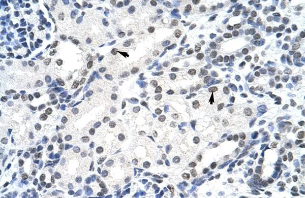
Immunohistochemical staining of human skeletal muscle shows strong positivity in nucleoli in myocytes.

Immunohistochemical staining of human skeletal muscle shows strong positivity in nucleoli in myocytes.
Anti-SURF6 Antibody
HPA023608
ApplicationsImmunoCytoChemistry, ImmunoHistoChemistry
Product group Antibodies
ReactivityHuman
TargetSURF6
Overview
- SupplierAtlas Antibodies
- Product NameAnti-SURF6 Antibody
- Delivery Days Customer4
- ApplicationsImmunoCytoChemistry, ImmunoHistoChemistry
- CertificationResearch Use Only
- ClonalityPolyclonal
- ConjugateUnconjugated
- Gene ID6838
- Target nameSURF6
- Target descriptionsurfeit 6
- Target synonymsRRP14, surfeit locus protein 6
- HostRabbit
- IsotypeIgG
- Protein IDO75683
- Protein NameSurfeit locus protein 6
- Scientific DescriptionRecombinant Protein Epitope Signature Tag (PrEST) antigen sequence
- ReactivityHuman
- Storage Instruction-20°C,2°C to 8°C
- UNSPSC41116161
Datasheet
MSDS
Related products
Product group Antibodies
Anti-SURF6 (Center) Antibody102-23460
ApplicationsWestern Blot
TargetSURF6
- SizePrice
Product group Antibodies
SURF6 antibody, InternalGTX47336
ApplicationsWestern Blot, ImmunoHistoChemistry, ImmunoHistoChemistry Paraffin
ReactivityHuman
TargetSURF6
- SizePrice
Product group Antibodies

SURF6 Antibody (aa187-213)LS-C157114
ApplicationsWestern Blot
ReactivityHuman
TargetSURF6
- SizePrice
Product group Antibodies
Anti-SURF6 AntibodyHPA074622
ApplicationsWestern Blot, ImmunoCytoChemistry
ReactivityHuman
TargetSURF6
- SizePrice

Product group Antibodies
Anti-SURF6 Antibody Picoband(r)A11571-1-CARRIER-FREE
ApplicationsImmunoFluorescence, Western Blot, ELISA, ImmunoCytoChemistry
ReactivityHuman
TargetSURF6
- SizePrice