

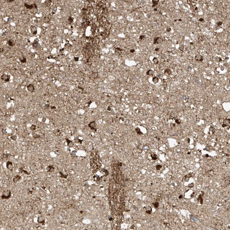
Immunohistochemical staining of human caudate shows strong cytoplasmic positivity in glial cells.
![Lane 1: Marker [kDa] 230, 130, 95, 72, 56, 36, 28, 17, 11. Lane 2: Human cell line RT-4](https://atlasantibodies.s3.amazonaws.com/images/wb/hpa031000-wb-1.jpg "Lane 1: Marker [kDa] 230, 130, 95, 72, 56, 36, 28, 17, 11. Lane 2: Human cell line RT-4")
Immunohistochemical staining of human caudate shows strong cytoplasmic positivity in glial cells.
Anti-TAGAP Antibody
HPA031000
ApplicationsWestern Blot, ImmunoHistoChemistry
Product group Antibodies
ReactivityHuman
TargetTAGAP
Overview
- SupplierAtlas Antibodies
- Product NameAnti-TAGAP Antibody
- Delivery Days Customer4
- ApplicationsWestern Blot, ImmunoHistoChemistry
- CertificationResearch Use Only
- ClonalityPolyclonal
- ConjugateUnconjugated
- Gene ID117289
- Target nameTAGAP
- Target descriptionT cell activation RhoGTPase activating protein
- Target synonymsARHGAP47, FKSG15, IDDM21, TAGAP1, T-cell activation Rho GTPase-activating protein
- HostRabbit
- IsotypeIgG
- Protein IDQ8N103
- Protein NameT-cell activation Rho GTPase-activating protein
- Scientific DescriptionRecombinant Protein Epitope Signature Tag (PrEST) antigen sequence
- ReactivityHuman
- Storage Instruction-20°C,2°C to 8°C
- UNSPSC41116161
Datasheet
MSDS
Related products
Product group Antibodies
Anti-TAGAP (Center) Antibody102-20676
ApplicationsFlow Cytometry, Western Blot, ImmunoHistoChemistry, ImmunoHistoChemistry Paraffin
TargetTAGAP
- SizePrice

Product group Antibodies
Anti-TAGAP AntibodyA100639
ApplicationsELISA, ImmunoHistoChemistry
ReactivityHuman
- SizePrice


Product group Antibodies
ApplicationsImmunoPrecipitation, Western Blot
ReactivityHuman, Mouse, Rat
TargetTAGAP
- SizePrice
Product group Antibodies

TAGAP AntibodyLS-C831979
ApplicationsWestern Blot, ELISA
ReactivityHuman, Mouse
TargetTAGAP
- SizePrice
Product group Antibodies
TAGAP Recombinant AntibodyBSM-62745R
ApplicationsImmunoPrecipitation, Western Blot
ReactivityHuman, Mouse, Rat
TargetTAGAP
- SizePrice
Product group Antibodies
TAGAP AntibodyCSB-PA006897
ApplicationsELISA, ImmunoHistoChemistry
ReactivityHuman
TargetTAGAP
- SizePrice
Product group Antibodies
TAGAP antibodyGTX87132
ApplicationsImmunoHistoChemistry, ImmunoHistoChemistry Paraffin
ReactivityHuman
TargetTAGAP
- SizePrice