


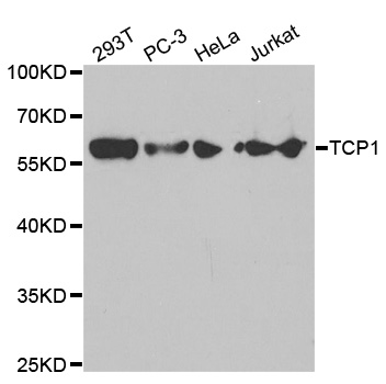
Figure 1. Western blot analysis of TCP1 using anti-TCP1 antibody (PB9826). Electrophoresis was performed on a 5-20% SDS-PAGE gel at 70V (Stacking gel) / 90V (Resolving gel) for 2-3 hours. The sample well of each lane was loaded with 30 ug of sample under reducing conditions. Lane 1: human A431 whole cell lysates, Lane 2: human Hela whole cell lysates, Lane 3: human 293T whole cell lysates, Lane 4: human MOLT4 whole cell lysates, Lane 5: human Jurkat whole cell lysates, Lane 6: human A549 whole cell lysates, Lane 7: human MCF-7 whole cell lysates, Lane 8: human U251 whole cell lysates. After electrophoresis, proteins were transferred to a nitrocellulose membrane at 150 mA for 50-90 minutes. Blocked the membrane with 5% non-fat milk/TBS for 1.5 hour at RT. The membrane was incubated with rabbit anti-TCP1 antigen affinity purified polyclonal antibody (Catalog # PB9826) at 0.5 microg/mL overnight at 4°C, then washed with TBS-0.1%Tween 3 times with 5 minutes each and probed with a goat anti-rabbit IgG-HRP secondary antibody at a dilution of 1:5000 for 1.5 hour at RT. The signal is developed using an Enhanced Chemiluminescent detection (ECL) kit (Catalog # EK1002) with Tanon 5200 system. A specific band was detected for TCP1 at approximately 60 kDa. The expected band size for TCP1 is at 60 kDa.
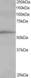
. Electrophoresis was performed on a 5-20% SDS-PAGE gel at 70V (Stacking gel) / 90V (Resolving gel) for 2-3 hours. The sample well of each lane was loaded with 30 ug of sample under reducing conditions. Lane 1: rat heart tissue lysates, Lane 2: rat ovary tissue lysates, Lane 3: rat brain tissue lysates, Lane 4: rat lung tissue lysates, Lane 5: mouse heart tissue lysates, Lane 6: mouse ovary tissue lysates, Lane 7: mouse brain tissue lysates, Lane 8: mouse lung tissue lysates. After electrophoresis, proteins were transferred to a nitrocellulose membrane at 150 mA for 50-90 minutes. Blocked the membrane with 5% non-fat milk/TBS for 1.5 hour at RT. The membrane was incubated with rabbit anti-TCP1 antigen affinity purified polyclonal antibody (Catalog # PB9826) at 0.5 microg/mL overnight at 4°C, then washed with TBS-0.1%Tween 3 times with 5 minutes each and probed with a goat anti-rabbit IgG-HRP secondary antibody at a dilution of 1:5000 for 1.5 hour at RT. The signal is developed using an Enhanced Chemiluminescent detection (ECL) kit (Catalog # EK1002) with Tanon 5200 system. A specific band was detected for TCP1 at approximately 60 kDa. The expected band size for TCP1 is at 60 kDa.")
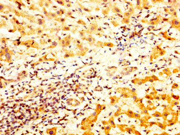
. TCP1 was detected in a paraffin-embedded section of human ovary cancer tissue. Heat mediated antigen retrieval was performed in EDTA buffer (pH 8.0, epitope retrieval solution). The tissue section was blocked with 10% goat serum. The tissue section was then incubated with 2 microg/ml rabbit anti-TCP1 Antibody (PB9826) overnight at 4°C. Peroxidase Conjugated Goat Anti-rabbit IgG was used as secondary antibody and incubated for 30 minutes at 37°C. The tissue section was developed using HRP Conjugated Rabbit IgG Super Vision Assay Kit (Catalog # SV0002) with DAB as the chromogen.")
. TCP1 was detected in a paraffin-embedded section of human testis tissue. Heat mediated antigen retrieval was performed in EDTA buffer (pH 8.0, epitope retrieval solution). The tissue section was blocked with 10% goat serum. The tissue section was then incubated with 2 microg/ml rabbit anti-TCP1 Antibody (PB9826) overnight at 4°C. Peroxidase Conjugated Goat Anti-rabbit IgG was used as secondary antibody and incubated for 30 minutes at 37°C. The tissue section was developed using HRP Conjugated Rabbit IgG Super Vision Assay Kit (Catalog # SV0002) with DAB as the chromogen.")
. TCP1 was detected in a paraffin-embedded section of mouse testis tissue. Heat mediated antigen retrieval was performed in EDTA buffer (pH 8.0, epitope retrieval solution). The tissue section was blocked with 10% goat serum. The tissue section was then incubated with 2 microg/ml rabbit anti-TCP1 Antibody (PB9826) overnight at 4°C. Peroxidase Conjugated Goat Anti-rabbit IgG was used as secondary antibody and incubated for 30 minutes at 37°C. The tissue section was developed using HRP Conjugated Rabbit IgG Super Vision Assay Kit (Catalog # SV0002) with DAB as the chromogen.")
. TCP1 was detected in a paraffin-embedded section of rat testis tissue. Heat mediated antigen retrieval was performed in EDTA buffer (pH 8.0, epitope retrieval solution). The tissue section was blocked with 10% goat serum. The tissue section was then incubated with 2 microg/ml rabbit anti-TCP1 Antibody (PB9826) overnight at 4°C. Peroxidase Conjugated Goat Anti-rabbit IgG was used as secondary antibody and incubated for 30 minutes at 37°C. The tissue section was developed using HRP Conjugated Rabbit IgG Super Vision Assay Kit (Catalog # SV0002) with DAB as the chromogen.")
. TCP1 was detected in immunocytochemical section of CACO-2 cells. Enzyme antigen retrieval was performed using IHC enzyme antigen retrieval reagent (AR0022) for 15 mins. The cells were blocked with 10% goat serum. And then incubated with 5microg/mL rabbit anti- TCP1 Antibody (PB9077) overnight at 4°C. DyLight®488 Conjugated Goat Anti-Rabbit IgG (BA1127) was used as secondary antibody at 1:500 dilution and incubated for 30 minutes at 37°C. The section was counterstained with DAPI. Visualize using a fluorescence microscope and filter sets appropriate for the label used.")
Figure 1. Western blot analysis of TCP1 using anti-TCP1 antibody (PB9826). Electrophoresis was performed on a 5-20% SDS-PAGE gel at 70V (Stacking gel) / 90V (Resolving gel) for 2-3 hours. The sample well of each lane was loaded with 30 ug of sample under reducing conditions. Lane 1: human A431 whole cell lysates, Lane 2: human Hela whole cell lysates, Lane 3: human 293T whole cell lysates, Lane 4: human MOLT4 whole cell lysates, Lane 5: human Jurkat whole cell lysates, Lane 6: human A549 whole cell lysates, Lane 7: human MCF-7 whole cell lysates, Lane 8: human U251 whole cell lysates. After electrophoresis, proteins were transferred to a nitrocellulose membrane at 150 mA for 50-90 minutes. Blocked the membrane with 5% non-fat milk/TBS for 1.5 hour at RT. The membrane was incubated with rabbit anti-TCP1 antigen affinity purified polyclonal antibody (Catalog # PB9826) at 0.5 microg/mL overnight at 4°C, then washed with TBS-0.1%Tween 3 times with 5 minutes each and probed with a goat anti-rabbit IgG-HRP secondary antibody at a dilution of 1:5000 for 1.5 hour at RT. The signal is developed using an Enhanced Chemiluminescent detection (ECL) kit (Catalog # EK1002) with Tanon 5200 system. A specific band was detected for TCP1 at approximately 60 kDa. The expected band size for TCP1 is at 60 kDa.
Anti-TCP1 alpha Antibody Picoband(r)
PB9826-CARRIER-FREE
ApplicationsImmunoFluorescence, Western Blot, ImmunoCytoChemistry, ImmunoHistoChemistry
Product group Antibodies
ReactivityBovine, Human, Mouse, Rat
TargetTCP1
Overview
- SupplierBoster Bio
- Product NameAnti-TCP1 alpha Antibody Picoband(r)
- Delivery Days Customer9
- Application Supplier NoteTested Species: In-house tested species with positive results. By Heat: Boiling the paraffin sections in 10mM citrate buffer, pH6.0, for 20mins is required for the staining of formalin/paraffin sections. Other applications have not been tested. Optimal dilutions should be determined by end users.
- ApplicationsImmunoFluorescence, Western Blot, ImmunoCytoChemistry, ImmunoHistoChemistry
- CertificationResearch Use Only
- ClonalityPolyclonal
- Concentration500 ug/ml
- Gene ID6950
- Target nameTCP1
- Target descriptiont-complex 1
- Target synonymsCCT-alpha, CCT1, CCTa, D6S230E, IDDPMGS, TCP-1-alpha, T-complex protein 1 subunit alpha, T-complex protein 1, alpha subunit, chaperonin containing T-complex polypeptide 1 subunit 1, t-complex 1 protein, tailless complex polypeptide 1
- HostRabbit
- IsotypeIgG
- Protein IDP17987
- Protein NameT-complex protein 1 subunit alpha
- Scientific DescriptionBoster Bio Anti-TCP1 alpha Antibody Picoband® catalog # PB9826. Tested in IF, IHC, ICC, WB applications. This antibody reacts with Human, Mouse, Rat. The brand Picoband indicates this is a premium antibody that guarantees superior quality, high affinity, and strong signals with minimal background in Western blot applications. Only our best-performing antibodies are designated as Picoband, ensuring unmatched performance.
- ReactivityBovine, Human, Mouse, Rat
- Storage Instruction-20°C,2°C to 8°C
- UNSPSC12352203
Related products
Product group Antibodies
Anti-TCP1 Antibody144-60306
ApplicationsImmunoFluorescence, Western Blot
ReactivityHuman, Mouse
TargetTCP1
- SizePrice
Product group Antibodies
Anti-TCP1 AntibodyA30532
ApplicationsImmunoFluorescence, Western Blot, ImmunoHistoChemistry
ReactivityHuman, Mouse, Rat
- SizePrice
Product group Antibodies
TCP1 alpha Recombinant Antibody, AbBy Fluor-647 ConjugatedBSM-62061R-BF647
ApplicationsImmunoFluorescence, Western Blot
ReactivityHuman
TargetTCP1
- SizePrice
Product group Antibodies

Goat anti-TCP1EB06277
ApplicationsFlow Cytometry, ImmunoFluorescence, Western Blot, ELISA, ImmunoHistoChemistry
ReactivityHuman
TargetTCP1
- SizePrice
Product group Antibodies
TCP1 AntibodyCSB-PA023320LA01HU
ApplicationsImmunoFluorescence, ELISA, ImmunoHistoChemistry
ReactivityHuman
TargetTCP1
- SizePrice
Product group Antibodies
TCP1 alpha antibody, C-termGTX10180
ApplicationsWestern Blot, ImmunoHistoChemistry, ImmunoHistoChemistry Paraffin
ReactivityHuman
TargetTCP1
- SizePrice

Product group Antibodies
Anti-TCP1 AntibodyHPA027337
ApplicationsWestern Blot, ImmunoHistoChemistry
ReactivityHuman, Mouse, Rat
TargetTCP1
- SizePrice
Product group Antibodies

TCP1 AntibodyLS-C761303
ApplicationsWestern Blot
ReactivityHuman
TargetTCP1
- SizePrice
Product group Antibodies
Anti-TCP1 AntibodyCAB13364
ApplicationsWestern Blot, ELISA, ImmunoHistoChemistry, ImmunoHistoChemistry Paraffin
ReactivityHuman
TargetTCP1
- SizePrice