


Immunohistochemical staining of human cerebellum shows strong nuclear positivity in Purkinje cells.

Immunohistochemical staining of human cerebellum shows strong nuclear positivity in Purkinje cells.
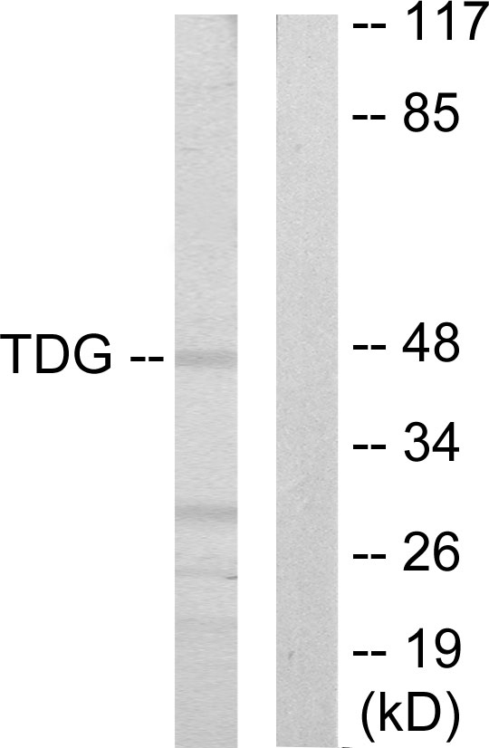
Anti-TDG Antibody
HPA052263
ApplicationsImmunoCytoChemistry, ImmunoHistoChemistry
Product group Antibodies
ReactivityHuman
TargetTDG
Overview
- SupplierAtlas Antibodies
- Product NameAnti-TDG Antibody
- Delivery Days Customer4
- ApplicationsImmunoCytoChemistry, ImmunoHistoChemistry
- CertificationResearch Use Only
- ClonalityPolyclonal
- ConjugateUnconjugated
- Gene ID6996
- Target nameTDG
- Target descriptionthymine DNA glycosylase
- Target synonymshTDG, G/T mismatch-specific thymine DNA glycosylase
- HostRabbit
- IsotypeIgG
- Protein IDQ13569
- Protein NameG/T mismatch-specific thymine DNA glycosylase
- Scientific DescriptionRecombinant Protein Epitope Signature Tag (PrEST) antigen sequence
- ReactivityHuman
- Storage Instruction-20°C,2°C to 8°C
- UNSPSC41116161
Datasheet
MSDS
Related products

Product group Antibodies
Anti-TDG Antibody Picoband(r)A15317-1-CARRIER-FREE
ApplicationsFlow Cytometry, Western Blot, ELISA
ReactivityHuman, Monkey
TargetTDG
- SizePrice
Product group Antibodies
Anti-TDG Antibody144-05756
ApplicationsWestern Blot
ReactivityHuman, Mouse, Rat
TargetTDG
- SizePrice
Product group Antibodies
Anti-TDG AntibodyA98428
ApplicationsWestern Blot, ELISA
ReactivityHuman, Mouse, Rat
- SizePrice
Product group Antibodies

TDG / Thymine DNA Glycosylase AntibodyLS-C748418
ApplicationsImmunoFluorescence, Western Blot
ReactivityHuman, Mouse, Rat
TargetTDG
- SizePrice
Product group Antibodies
TDG AntibodyCSB-PA050229
ApplicationsImmunoFluorescence, Western Blot, ELISA, ImmunoHistoChemistry
ReactivityHuman, Mouse, Rat
TargetTDG
- SizePrice

Product group Antibodies
TDG antibodyGTX110473
ApplicationsImmunoFluorescence, Western Blot, ImmunoCytoChemistry
ReactivityHuman, Mouse
TargetTDG
- SizePrice