


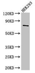
Figure 1. Western blot analysis of TDRD3 using anti-TDRD3 antibody (A08978). Electrophoresis was performed on a 5-20% SDS-PAGE gel at 70V (Stacking gel) / 90V (Resolving gel) for 2-3 hours. The sample well of each lane was loaded with 50ug of sample under reducing conditions. Lane 1: human placenta tissue lysates, Lane 2: human HL-60 whole cell lysates, Lane 3: human Hela whole cell lysates, Lane 4: human PC-3 whole cell lysates, Lane 5: human HEK293 whole cell lysates, Lane 6: human A549 whole cell lysates, Lane 7: human A431 whole cell lysates. After Electrophoresis, proteins were transferred to a Nitrocellulose membrane at 150mA for 50-90 minutes. Blocked the membrane with 5% Non-fat Milk/ TBS for 1.5 hour at RT. The membrane was incubated with rabbit anti-TDRD3 antigen affinity purified polyclonal antibody (Catalog # A08978) at 0.5 microg/mL overnight at 4°C, then washed with TBS-0.1%Tween 3 times with 5 minutes each and probed with a goat anti-rabbit IgG-HRP secondary antibody at a dilution of 1:10000 for 1.5 hour at RT. The signal is developed using an Enhanced Chemiluminescent detection (ECL) kit (Catalog # EK1002) with Tanon 5200 system. Specific bands were detected for TDRD3 at approximately 65-73KD. The expected band size for TDRD3 is at 73KD.
. Electrophoresis was performed on a 5-20% SDS-PAGE gel at 70V (Stacking gel) / 90V (Resolving gel) for 2-3 hours. The sample well of each lane was loaded with 50ug of sample under reducing conditions. Lane 1: rat brain tissues lysates Lane 2: rat liver tissue lysates Lane 3: mouse brain tissue lysates Lane 4: mouse lung tissue lysates Lane 5: mouse liver tissue lysates Lane 6: mouse NIH3T3 whole cell lysates After Electrophoresis, proteins were transferred to a Nitrocellulose membrane at 150mA for 50-90 minutes. Blocked the membrane with 5% Non-fat Milk/ TBS for 1.5 hour at RT. The membrane was incubated with rabbit anti-TDRD3 antigen affinity purified polyclonal antibody (Catalog # A08978) at 0.5 microg/mL overnight at 4°C, then washed with TBS-0.1%Tween 3 times with 5 minutes each and probed with a goat anti-rabbit IgG-HRP secondary antibody at a dilution of 1:10000 for 1.5 hour at RT. The signal is developed using an Enhanced Chemiluminescent detection (ECL) kit (Catalog # EK1002) with Tanon 5200 system. A specific band was detected for TDRD3 at approximately 65-73KD. The expected band size for TDRD3 is at 73KD.")
. TDRD3 was detected in paraffin-embedded section of human colon cancer tissue. Heat mediated antigen retrieval was performed in citrate buffer (pH6, epitope retrieval solution) for 20 mins. The tissue section was blocked with 10% goat serum. The tissue section was then incubated with 1microg/ml rabbit anti-TDRD3 Antibody (A08978) overnight at 4°C. Biotinylated goat anti-rabbit IgG was used as secondary antibody and incubated for 30 minutes at 37°C. The tissue section was developed using Strepavidin-Biotin-Complex (SABC)(Catalog # SA1022) with DAB as the chromogen.")
. TDRD3 was detected in paraffin-embedded section of mouse small intestine tissue. Heat mediated antigen retrieval was performed in citrate buffer (pH6, epitope retrieval solution) for 20 mins. The tissue section was blocked with 10% goat serum. The tissue section was then incubated with 1microg/ml rabbit anti-TDRD3 Antibody (A08978) overnight at 4°C. Biotinylated goat anti-rabbit IgG was used as secondary antibody and incubated for 30 minutes at 37°C. The tissue section was developed using Strepavidin-Biotin-Complex (SABC)(Catalog # SA1022) with DAB as the chromogen.")
. TDRD3 was detected in paraffin-embedded section of rat small intestine tissue. Heat mediated antigen retrieval was performed in citrate buffer (pH6, epitope retrieval solution) for 20 mins. The tissue section was blocked with 10% goat serum. The tissue section was then incubated with 1microg/ml rabbit anti-TDRD3 Antibody (A08978) overnight at 4°C. Biotinylated goat anti-rabbit IgG was used as secondary antibody and incubated for 30 minutes at 37°C. The tissue section was developed using Strepavidin-Biotin-Complex (SABC)(Catalog # SA1022) with DAB as the chromogen.")
 TDRD3 was detected in paraffin-embedded section of human intestinal cancer tissues. Heat mediated antigen retrieval was performed in citrate buffer (pH6, epitope retrieval solution ) for 20 mins. The tissue section was blocked with 10% goat serum. The tissue section was then incubated with 1microg/mL rabbit anti-TDRD3 Antibody (A08978) overnight at 4°C. Biotin conjugated goat anti-rabbit IgG (BA1003) was used as secondary antibody and incubated for 30 minutes at 37°C. The tissue section was developed using DyLight®550 Conjugated Avidin (BA1134). The section was counterstained with DAPI. Visualize using a fluorescence microscope and filter sets appropriate for the label used.")
Figure 1. Western blot analysis of TDRD3 using anti-TDRD3 antibody (A08978). Electrophoresis was performed on a 5-20% SDS-PAGE gel at 70V (Stacking gel) / 90V (Resolving gel) for 2-3 hours. The sample well of each lane was loaded with 50ug of sample under reducing conditions. Lane 1: human placenta tissue lysates, Lane 2: human HL-60 whole cell lysates, Lane 3: human Hela whole cell lysates, Lane 4: human PC-3 whole cell lysates, Lane 5: human HEK293 whole cell lysates, Lane 6: human A549 whole cell lysates, Lane 7: human A431 whole cell lysates. After Electrophoresis, proteins were transferred to a Nitrocellulose membrane at 150mA for 50-90 minutes. Blocked the membrane with 5% Non-fat Milk/ TBS for 1.5 hour at RT. The membrane was incubated with rabbit anti-TDRD3 antigen affinity purified polyclonal antibody (Catalog # A08978) at 0.5 microg/mL overnight at 4°C, then washed with TBS-0.1%Tween 3 times with 5 minutes each and probed with a goat anti-rabbit IgG-HRP secondary antibody at a dilution of 1:10000 for 1.5 hour at RT. The signal is developed using an Enhanced Chemiluminescent detection (ECL) kit (Catalog # EK1002) with Tanon 5200 system. Specific bands were detected for TDRD3 at approximately 65-73KD. The expected band size for TDRD3 is at 73KD.
Anti-TDRD3 Antibody Picoband(r)
A08978-CARRIER-FREE
ApplicationsImmunoFluorescence, Western Blot, ImmunoHistoChemistry
Product group Antibodies
ReactivityHuman, Mouse, Rat
TargetTDRD3
Overview
- SupplierBoster Bio
- Product NameAnti-TDRD3 Antibody Picoband(r)
- Delivery Days Customer9
- Application Supplier NoteTested Species: In-house tested species with positive results. By Heat: Boiling the paraffin sections in 10mM citrate buffer, pH6.0, for 20mins is required for the staining of formalin/paraffin sections. Other applications have not been tested. Optimal dilutions should be determined by end users.
- ApplicationsImmunoFluorescence, Western Blot, ImmunoHistoChemistry
- CertificationResearch Use Only
- ClonalityPolyclonal
- Concentration500 ug/ml
- Gene ID81550
- Target nameTDRD3
- Target descriptiontudor domain containing 3
- Target synonymstudor domain-containing protein 3
- HostRabbit
- IsotypeIgG
- Protein IDQ9H7E2
- Protein NameTudor domain-containing protein 3
- Scientific DescriptionBoster Bio Anti-TDRD3 Antibody Picoband® catalog # A08978. Tested in IF, IHC, WB applications. This antibody reacts with Human, Mouse, Rat. The brand Picoband indicates this is a premium antibody that guarantees superior quality, high affinity, and strong signals with minimal background in Western blot applications. Only our best-performing antibodies are designated as Picoband, ensuring unmatched performance.
- ReactivityHuman, Mouse, Rat
- Storage Instruction-20°C,2°C to 8°C
- UNSPSC12352203
Related products
Product group Antibodies

Anti-TDRD3 [Fab-8-1]Ab00408-1.1
ApplicationsELISA
ReactivityHuman
TargetTDRD3
- SizePrice
Product group Antibodies
Anti-TDRD3 Antibody144-66040
ApplicationsWestern Blot
ReactivityHuman
TargetTDRD3
- SizePrice
Product group Antibodies
TDRD3 Polyclonal AntibodyCAC14570
ApplicationsWestern Blot, ELISA, ImmunoHistoChemistry
TargetTDRD3
- SizePrice


Product group Antibodies
Anti-TDRD3 AntibodyHPA067153
ApplicationsImmunoCytoChemistry
ReactivityHuman
TargetTDRD3
- SizePrice
Product group Antibodies
TDRD3 AntibodyCSB-PA863996LA01HU
ApplicationsWestern Blot, ELISA, ImmunoHistoChemistry
ReactivityHuman
TargetTDRD3
- SizePrice
Product group Antibodies

TDRD3 AntibodyLS-C335623
ApplicationsWestern Blot
ReactivityHuman
TargetTDRD3
- SizePrice