


Immunofluorescent staining of human cell line U-251 MG shows localization to mitochondria.

Immunofluorescent staining of human cell line U-251 MG shows localization to mitochondria.
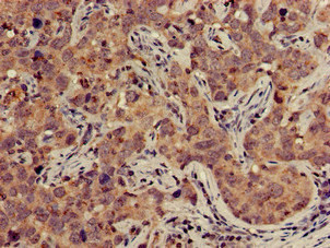
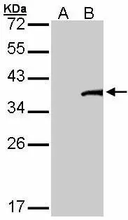
Anti-TFB2M Antibody
HPA028482
ApplicationsWestern Blot, ImmunoCytoChemistry
Product group Antibodies
ReactivityHuman
TargetTFB2M
Overview
- SupplierAtlas Antibodies
- Product NameAnti-TFB2M Antibody
- Delivery Days Customer4
- ApplicationsWestern Blot, ImmunoCytoChemistry
- CertificationResearch Use Only
- ClonalityPolyclonal
- ConjugateUnconjugated
- Gene ID64216
- Target nameTFB2M
- Target descriptiontranscription factor B2, mitochondrial
- Target synonymsHkp1, h-mtTFB, h-mtTFB2, hTFB2M, mtTFB2, dimethyladenosine transferase 2, mitochondrial, HCV NS5A-transactivated protein 5, S-adenosylmethionine-6-N', N'-adenosyl(rRNA) dimethyltransferase 2, hepatitis C virus NS5A-transactivated protein 5, mitochondrial 12S rRNA dimethylase 2, mitochondrial transcription factor B2
- HostRabbit
- IsotypeIgG
- Protein IDQ9H5Q4
- Protein NameDimethyladenosine transferase 2, mitochondrial
- Scientific DescriptionRecombinant Protein Epitope Signature Tag (PrEST) antigen sequence
- ReactivityHuman
- Storage Instruction-20°C,2°C to 8°C
- UNSPSC41116161
Datasheet
MSDS
Related products

Product group Antibodies
Anti-TFB2M Antibody Picoband(r)A05679-2-CARRIER-FREE
ApplicationsFlow Cytometry, Western Blot, ELISA
ReactivityHuman
TargetTFB2M
- SizePrice
Product group Antibodies

TFB2M AntibodyLS-C749899
ApplicationsWestern Blot
ReactivityHuman, Mouse
TargetTFB2M
- SizePrice
Product group Antibodies

Goat anti-TFB2MEB05656
ApplicationsELISA, ImmunoHistoChemistry
ReactivityHuman
TargetTFB2M
- SizePrice
Product group Antibodies
TFB2M AntibodyCSB-PA867154LA01HU
ApplicationsELISA, ImmunoHistoChemistry
ReactivityHuman
TargetTFB2M
- SizePrice

Product group Antibodies
Anti-TFB2M AntibodyHPA028554
ApplicationsImmunoCytoChemistry
ReactivityHuman
TargetTFB2M
- SizePrice

Product group Antibodies
Anti-TFB2M AntibodyHPA030265
ApplicationsWestern Blot, ImmunoHistoChemistry
ReactivityHuman
TargetTFB2M
- SizePrice
Product group Antibodies
TFB2M antibody [C3], C-termGTX107714
ApplicationsWestern Blot
ReactivityHuman
TargetTFB2M
- SizePrice