


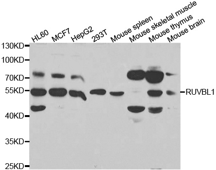
Figure 1. Western blot analysis of TIP49A/RUVBL1 using anti-TIP49A/RUVBL1 antibody (A02049-2). Electrophoresis was performed on a 5-20% SDS-PAGE gel at 70V (Stacking gel) / 90V (Resolving gel) for 2-3 hours. The sample well of each lane was loaded with 30 ug of sample under reducing conditions. Lane 1: human K562 whole cell lysates, Lane 2: human 293T whole cell lysates, Lane 3: human HT1080 whole cell lysates, Lane 4: monkey COS-7 whole cell lysates, Lane 5: human MCF-7 whole cell lysates, Lane 6: human Daudi whole cell lysates, Lane 7: human MOLT-4 whole cell lysates, Lane 8: human HEL whole cell lysates, Lane 9: rat testis tissue lysates, Lane 10: rat C6 whole cell lysates, Lane 11: mouse testis tissue lysates, Lane 12: mouse NIH/3T3 whole cell lysates. red to a nitrocellulose membrane at 150 mA for 50-90 minutes. Blocked the membrane with 5% non-fat milk/TBS for 1.5 hour at RT. The membrane was incubated with rabbit anti-TIP49A/RUVBL1 antigen affinity purified polyclonal antibody (Catalog # A02049-2) at 0.25 microg/mL overnight at 4°C, then washed with TBS-0.1%Tween 3 times with 5 minutes each and probed with a goat anti-rabbit IgG-HRP secondary antibody at a dilution of 1:5000 for 1.5 hour at RT. The signal is developed using an Enhanced Chemiluminescent detection (ECL) kit (Catalog # EK1002) with Tanon 5200 system. A specific band was detected for TIP49A/RUVBL1 at approximately 54 kDa. The expected band size for TIP49A/RUVBL1 is at 50 kDa.
. TIP49A/RUVBL1 was detected in a paraffin-embedded section of human colorectal adenocarcinoma tissue. Heat mediated antigen retrieval was performed in EDTA buffer (pH 8.0, epitope retrieval solution). The tissue section was blocked with 10% goat serum. The tissue section was then incubated with 2 microg/ml rabbit anti-TIP49A/RUVBL1 Antibody (A02049-2) overnight at 4°C. Peroxidase Conjugated Goat Anti-rabbit IgG was used as secondary antibody and incubated for 30 minutes at 37°C. The tissue section was developed using HRP Conjugated Rabbit IgG Super Vision Assay Kit (Catalog # SV0002) with DAB as the chromogen.")
. TIP49A/RUVBL1 was detected in a paraffin-embedded section of human thyroid cancer tissue. Heat mediated antigen retrieval was performed in EDTA buffer (pH 8.0, epitope retrieval solution). The tissue section was blocked with 10% goat serum. The tissue section was then incubated with 2 microg/ml rabbit anti-TIP49A/RUVBL1 Antibody (A02049-2) overnight at 4°C. Peroxidase Conjugated Goat Anti-rabbit IgG was used as secondary antibody and incubated for 30 minutes at 37°C. The tissue section was developed using HRP Conjugated Rabbit IgG Super Vision Assay Kit (Catalog # SV0002) with DAB as the chromogen.")
. TIP49A/RUVBL1 was detected in a paraffin-embedded section of human lung adenocarcinoma tissue. Heat mediated antigen retrieval was performed in EDTA buffer (pH 8.0, epitope retrieval solution). The tissue section was blocked with 10% goat serum. The tissue section was then incubated with 2 microg/ml rabbit anti-TIP49A/RUVBL1 Antibody (A02049-2) overnight at 4°C. Peroxidase Conjugated Goat Anti-rabbit IgG was used as secondary antibody and incubated for 30 minutes at 37°C. The tissue section was developed using HRP Conjugated Rabbit IgG Super Vision Assay Kit (Catalog # SV0002) with DAB as the chromogen.")
. TIP49A/RUVBL1 was detected in a paraffin-embedded section of human spleen tissue. Heat mediated antigen retrieval was performed in EDTA buffer (pH 8.0, epitope retrieval solution). The tissue section was blocked with 10% goat serum. The tissue section was then incubated with 2 microg/ml rabbit anti-TIP49A/RUVBL1 Antibody (A02049-2) overnight at 4°C. Peroxidase Conjugated Goat Anti-rabbit IgG was used as secondary antibody and incubated for 30 minutes at 37°C. The tissue section was developed using HRP Conjugated Rabbit IgG Super Vision Assay Kit (Catalog # SV0002) with DAB as the chromogen.")
. TIP49A/RUVBL1 was detected in a paraffin-embedded section of human tonsil tissue. Heat mediated antigen retrieval was performed in EDTA buffer (pH 8.0, epitope retrieval solution). The tissue section was blocked with 10% goat serum. The tissue section was then incubated with 2 microg/ml rabbit anti-TIP49A/RUVBL1 Antibody (A02049-2) overnight at 4°C. Peroxidase Conjugated Goat Anti-rabbit IgG was used as secondary antibody and incubated for 30 minutes at 37°C. The tissue section was developed using HRP Conjugated Rabbit IgG Super Vision Assay Kit (Catalog # SV0002) with DAB as the chromogen.")
. TIP49A/RUVBL1 was detected in a paraffin-embedded section of human ovarian cancer tissue. Heat mediated antigen retrieval was performed in EDTA buffer (pH 8.0, epitope retrieval solution). The tissue section was blocked with 10% goat serum. The tissue section was then incubated with 2 microg/ml rabbit anti-TIP49A/RUVBL1 Antibody (A02049-2) overnight at 4°C. Peroxidase Conjugated Goat Anti-rabbit IgG was used as secondary antibody and incubated for 30 minutes at 37°C. The tissue section was developed using HRP Conjugated Rabbit IgG Super Vision Assay Kit (Catalog # SV0002) with DAB as the chromogen.")
. TIP49A/RUVBL1 was detected in an immunocytochemical section of A549 cells. Enzyme antigen retrieval was performed using IHC enzyme antigen retrieval reagent (AR0022) for 15 mins. The cells were blocked with 10% goat serum. And then incubated with 5 microg/mL rabbit anti-TIP49A/RUVBL1 Antibody (A02049-2) overnight at 4°C. DyLight®488 Conjugated Goat Anti-Rabbit IgG (BA1127) was used as secondary antibody at 1:100 dilution and incubated for 30 minutes at 37°C. The section was counterstained with DAPI. Visualize using a fluorescence microscope and filter sets appropriate for the label used.")
. TIP49A/RUVBL1 was detected in a paraffin-embedded section of human colon cancer tissue. Heat mediated antigen retrieval was performed in EDTA buffer (pH 8.0, epitope retrieval solution). The tissue section was blocked with 10% goat serum. The tissue section was then incubated with 5 microg/mL rabbit anti-TIP49A/RUVBL1 Antibody (A02049-2) overnight at 4°C. Cy3 Conjugated Goat Anti-Rabbit IgG (BA1032) was used as secondary antibody at 1:500 dilution and incubated for 30 minutes at 37°C. The section was counterstained with DAPI. Visualize using a fluorescence microscope and filter sets appropriate for the label used.")
. Overlay histogram showing Hela cells stained with A02049-2 (Blue line). To facilitate intracellular staining, cells were fixed with 4% paraformaldehyde and permeabilized with permeabilization buffer. The cells were blocked with 10% normal goat serum. And then incubated with rabbit anti-TIP49A/RUVBL1 Antibody (A02049-2, 1 microg/1x106 cells) for 30 min at 20°C. DyLight®488 conjugated goat anti-rabbit IgG (BA1127, 5-10 microg/1x106 cells) was used as secondary antibody for 30 minutes at 20°C. Isotype control antibody (Green line) was rabbit IgG (1 microg/1x106) used under the same conditions. Unlabelled sample without incubation with primary antibody and secondary antibody (Red line) was used as a blank control.")
Figure 1. Western blot analysis of TIP49A/RUVBL1 using anti-TIP49A/RUVBL1 antibody (A02049-2). Electrophoresis was performed on a 5-20% SDS-PAGE gel at 70V (Stacking gel) / 90V (Resolving gel) for 2-3 hours. The sample well of each lane was loaded with 30 ug of sample under reducing conditions. Lane 1: human K562 whole cell lysates, Lane 2: human 293T whole cell lysates, Lane 3: human HT1080 whole cell lysates, Lane 4: monkey COS-7 whole cell lysates, Lane 5: human MCF-7 whole cell lysates, Lane 6: human Daudi whole cell lysates, Lane 7: human MOLT-4 whole cell lysates, Lane 8: human HEL whole cell lysates, Lane 9: rat testis tissue lysates, Lane 10: rat C6 whole cell lysates, Lane 11: mouse testis tissue lysates, Lane 12: mouse NIH/3T3 whole cell lysates. red to a nitrocellulose membrane at 150 mA for 50-90 minutes. Blocked the membrane with 5% non-fat milk/TBS for 1.5 hour at RT. The membrane was incubated with rabbit anti-TIP49A/RUVBL1 antigen affinity purified polyclonal antibody (Catalog # A02049-2) at 0.25 microg/mL overnight at 4°C, then washed with TBS-0.1%Tween 3 times with 5 minutes each and probed with a goat anti-rabbit IgG-HRP secondary antibody at a dilution of 1:5000 for 1.5 hour at RT. The signal is developed using an Enhanced Chemiluminescent detection (ECL) kit (Catalog # EK1002) with Tanon 5200 system. A specific band was detected for TIP49A/RUVBL1 at approximately 54 kDa. The expected band size for TIP49A/RUVBL1 is at 50 kDa.
Anti-TIP49A/RUVBL1 Antibody Picoband(r)
A02049-2-CARRIER-FREE
ApplicationsFlow Cytometry, ImmunoFluorescence, Western Blot, ELISA, ImmunoCytoChemistry, ImmunoHistoChemistry
Product group Antibodies
ReactivityHuman, Monkey, Mouse, Rat
TargetRUVBL1
Overview
- SupplierBoster Bio
- Product NameAnti-TIP49A/RUVBL1 Antibody Picoband(r)
- Delivery Days Customer9
- ApplicationsFlow Cytometry, ImmunoFluorescence, Western Blot, ELISA, ImmunoCytoChemistry, ImmunoHistoChemistry
- CertificationResearch Use Only
- ClonalityPolyclonal
- Concentration500 ug/ml
- Gene ID8607
- Target nameRUVBL1
- Target descriptionRuvB like AAA ATPase 1
- Target synonymsECP-54, ECP54, INO80H, NMP 238, NMP238, PONTIN, Pontin52, RVB1, TIH1, TIP49, TIP49A, ruvB-like 1, 49 kDa TATA box-binding protein-interacting protein, 49 kDa TBP-interacting protein, 54 kDa erythrocyte cytosolic protein, INO80 complex subunit H, RuvB (E coli homolog)-like 1, RuvB-like AAA ATPase, TAP54-alpha, TATA binding protein interacting protein 49 kDa, TIP60-associated protein 54-alpha, epididymis secretory sperm binding protein, nuclear matrix protein 238, pontin 52
- HostRabbit
- IsotypeIgG
- Protein IDQ9Y265
- Protein NameRuvB-like 1
- Scientific DescriptionBoster Bio Anti-TIP49A/RUVBL1 Antibody Picoband® catalog # A02049-2. Tested in ELISA, Flow Cytometry, IF, IHC, ICC, WB applications. This antibody reacts with Human, Monkey, Mouse, Rat. The brand Picoband indicates this is a premium antibody that guarantees superior quality, high affinity, and strong signals with minimal background in Western blot applications. Only our best-performing antibodies are designated as Picoband, ensuring unmatched performance.
- ReactivityHuman, Monkey, Mouse, Rat
- Storage Instruction-20°C,2°C to 8°C
- UNSPSC12352203
Related products
Product group Antibodies
Anti-RUVBL1 AntibodyA31006
ApplicationsImmunoFluorescence, Western Blot, ImmunoHistoChemistry
ReactivityHuman, Mouse, Rat
- SizePrice
Product group Antibodies
Anti-RUVBL1 Antibody144-05723
ApplicationsImmunoFluorescence, Western Blot, ImmunoHistoChemistry
ReactivityHuman, Mouse
TargetRUVBL1
- SizePrice
Product group Antibodies

TIP49 / RUVBL1 AntibodyLS-C830521
ApplicationsWestern Blot, ELISA, ImmunoHistoChemistry
ReactivityHuman, Mouse, Rat
TargetRUVBL1
- SizePrice
Product group Antibodies
RUVBL1 Polyclonal AntibodyBS-19936R
ApplicationsImmunoFluorescence, Western Blot, ELISA, ImmunoCytoChemistry, ImmunoHistoChemistry, ImmunoHistoChemistry Frozen, ImmunoHistoChemistry Paraffin
ReactivityBovine, Canine, Chicken, Equine, Human, Mouse, Porcine, Rabbit, Rat, Sheep, Zebra Fish
TargetRUVBL1
- SizePrice
Product group Antibodies
RUVBL1 AntibodyCSB-PA896483LA01HU
ApplicationsImmunoFluorescence, Western Blot, ELISA, ImmunoHistoChemistry
ReactivityHuman
TargetRUVBL1
- SizePrice
Product group Antibodies
RUVBL1 Polyclonal AntibodyCAC14509
ApplicationsImmunoFluorescence, Western Blot, ELISA, ImmunoHistoChemistry
TargetRUVBL1
- SizePrice

Product group Antibodies
Anti-RUVBL1 AntibodyHPA019947
ApplicationsWestern Blot, ImmunoCytoChemistry, ImmunoHistoChemistry
ReactivityHuman
TargetRUVBL1
- SizePrice
Product group Antibodies
RUVBL1 antibodyGTX109494
ApplicationsImmunoPrecipitation, Western Blot
ReactivityHuman
TargetRUVBL1
- SizePrice