


Immunohistochemical staining of human caudate shows strong nuclear and cytoplasmic positivity in neuronal cells.
Immunohistochemical staining of human caudate shows strong nuclear and cytoplasmic positivity in neuronal cells.
Anti-TNK2 Antibody
HPA041954
ApplicationsImmunoHistoChemistry
Product group Antibodies
ReactivityHuman
TargetTNK2
Overview
- SupplierAtlas Antibodies
- Product NameAnti-TNK2 Antibody
- Delivery Days Customer4
- ApplicationsImmunoHistoChemistry
- CertificationResearch Use Only
- ClonalityPolyclonal
- ConjugateUnconjugated
- Gene ID10188
- Target nameTNK2
- Target descriptiontyrosine kinase non receptor 2
- Target synonymsACK, ACK-1, ACK1, p21cdc42Hs, activated CDC42 kinase 1, activated Cdc42-associated kinase 1, activated p21cdc42Hs kinase, tyrosine kinase non-receptor protein 2
- HostRabbit
- IsotypeIgG
- Protein IDQ07912
- Protein NameActivated CDC42 kinase 1
- Scientific DescriptionRecombinant Protein Epitope Signature Tag (PrEST) antigen sequence
- ReactivityHuman
- Storage Instruction-20°C,2°C to 8°C
- UNSPSC41116161
Datasheet
MSDS
Related products
Product group Antibodies
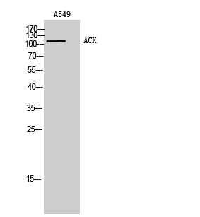
Anti-ACK1 AntibodyA99476
ApplicationsWestern Blot, ELISA
ReactivityHuman, Mouse
- SizePrice

Product group Antibodies
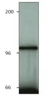
Anti-ACK1/TNK2 Antibody Picoband(r)A02334-1-CARRIER-FREE
ApplicationsFlow Cytometry, Western Blot, ELISA
ReactivityHuman
TargetTNK2
- SizePrice
Product group Antibodies
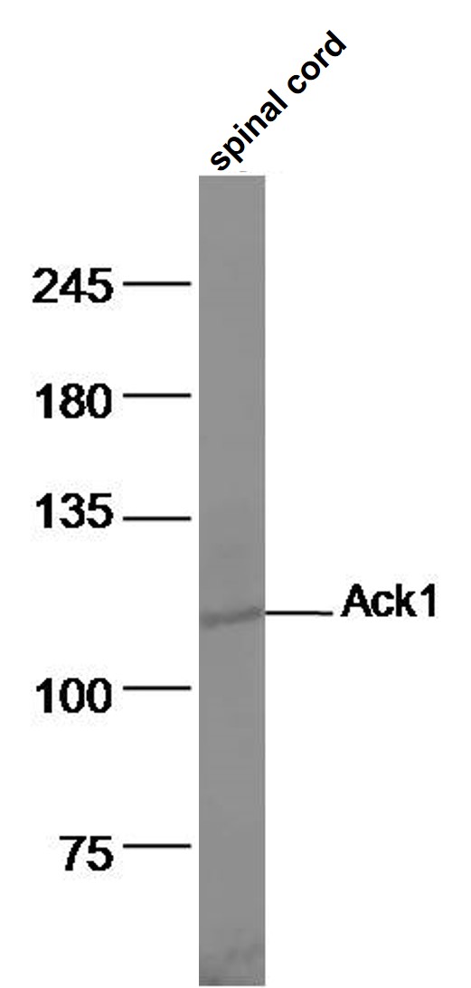
Ack1 Polyclonal AntibodyBS-1227R
ApplicationsImmunoFluorescence, Western Blot, ELISA, ImmunoCytoChemistry, ImmunoHistoChemistry, ImmunoHistoChemistry Frozen, ImmunoHistoChemistry Paraffin
ReactivityBovine, Canine, Chicken, Human, Mouse, Porcine, Rat
TargetTNK2
- SizePrice
Product group Antibodies
Phospho-TNK2 (Y284) AntibodyCSB-PA006413
ApplicationsImmunoFluorescence, Western Blot, ELISA, ImmunoHistoChemistry
ReactivityHuman, Mouse
TargetTNK2
- SizePrice
Product group Antibodies
ApplicationsImmunoPrecipitation, Western Blot, ImmunoCytoChemistry, ImmunoHistoChemistry
TargetTNK2
- SizePrice
Product group Antibodies

TNK2 / ACK1 AntibodyLS-C402095
ApplicationsWestern Blot, ELISA, ImmunoHistoChemistry
ReactivityHuman, Mouse, Rat
TargetTNK2
- SizePrice
Product group Antibodies
ACK1 antibodyGTX14776
ApplicationsImmunoPrecipitation, Western Blot, ELISA
ReactivityHuman, Mouse, Rat
TargetTNK2
- SizePrice