


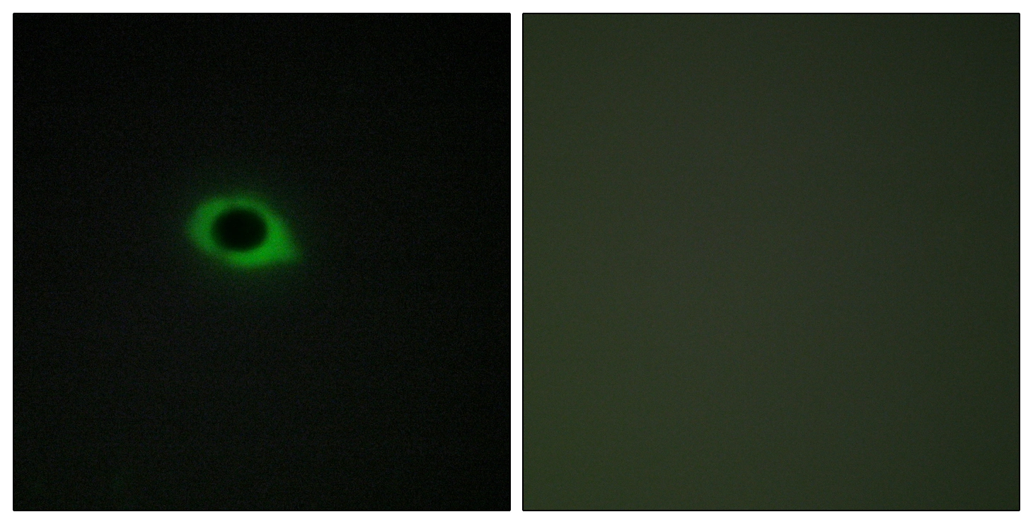
Immunohistochemical staining of human kidney shows strong cytoplasmic positivity in cells in tubules and cells in glomeruli.

Immunohistochemical staining of human kidney shows strong cytoplasmic positivity in cells in tubules and cells in glomeruli.
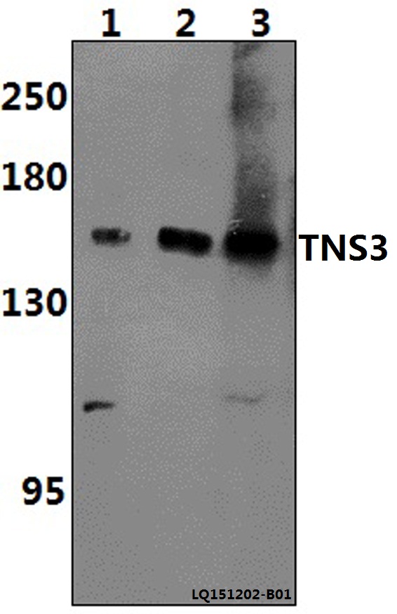
Anti-TNS3 Antibody
HPA056015
ApplicationsImmunoCytoChemistry, ImmunoHistoChemistry
Product group Antibodies
ReactivityHuman
TargetTNS3
Overview
- SupplierAtlas Antibodies
- Product NameAnti-TNS3 Antibody
- Delivery Days Customer4
- ApplicationsImmunoCytoChemistry, ImmunoHistoChemistry
- CertificationResearch Use Only
- ClonalityPolyclonal
- ConjugateUnconjugated
- Gene ID64759
- Target nameTNS3
- Target descriptiontensin 3
- Target synonymsTEM6, TENS1, tensin-3, tensin-like SH2 domain containing 1, tensin-like SH2 domain-containing protein 1, thyroid specific PTB domain protein, tumor endothelial marker 6
- HostRabbit
- IsotypeIgG
- Protein IDQ68CZ2
- Protein NameTensin-3
- Scientific DescriptionRecombinant Protein Epitope Signature Tag (PrEST) antigen sequence
- ReactivityHuman
- Storage Instruction-20°C,2°C to 8°C
- UNSPSC41116161
Datasheet
MSDS
Related products
Product group Antibodies
Anti-TNS3 AntibodyA29284
ApplicationsWestern Blot
ReactivityHuman, Mouse, Rat
- SizePrice

Product group Antibodies
Anti-TNS3 Antibody Picoband(r)A07345-1-CARRIER-FREE
ApplicationsFlow Cytometry, ImmunoFluorescence, ImmunoPrecipitation, Western Blot, ELISA, ImmunoCytoChemistry, ImmunoHistoChemistry
ReactivityHuman
TargetTNS3
- SizePrice
Product group Antibodies
ApplicationsImmunoPrecipitation, Western Blot, ImmunoCytoChemistry, ImmunoHistoChemistry
ReactivityPorcine
TargetTNS3
- SizePrice
Product group Antibodies
TNS3 AntibodyCSB-PA050061
ApplicationsImmunoFluorescence, ELISA, ImmunoHistoChemistry
ReactivityHuman
TargetTNS3
- SizePrice
Product group Antibodies

TNS3 / Tensin 3 AntibodyLS-C409535
ApplicationsWestern Blot, ImmunoHistoChemistry
ReactivityHuman
TargetTNS3
- SizePrice
Product group Antibodies
TENS3 antibodyGTX87949
ApplicationsImmunoFluorescence, ImmunoCytoChemistry, ImmunoHistoChemistry, ImmunoHistoChemistry Paraffin
ReactivityHuman, Monkey
TargetTNS3
- SizePrice

Product group Antibodies
Anti-TNS3 AntibodyHPA055338
ApplicationsWestern Blot, ImmunoCytoChemistry, ImmunoHistoChemistry
ReactivityHuman
TargetTNS3
- SizePrice