

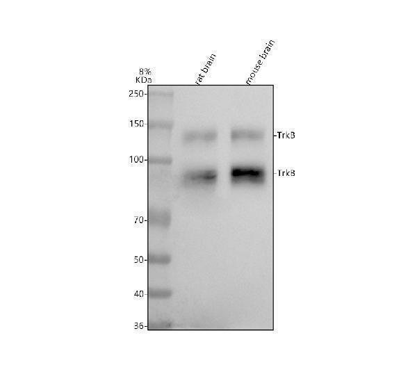
Figure 1. Western blot analysis of TrkB using anti-TrkB antibody (M01388-3). Electrophoresis was performed on a 5-20% SDS-PAGE gel at 70V (Stacking gel) / 90V (Resolving gel) for 2-3 hours. The sample well of each lane was loaded with 30 ug of sample under reducing conditions. Lane 1: rat brain tissue lysates, Lane 2: mouse brain tissue lysates. After electrophoresis, proteins were transferred to a nitrocellulose membrane at 150 mA for 50-90 minutes. Blocked the membrane with 5% non-fat milk/TBS for 1.5 hour at RT. The membrane was incubated with rabbit anti-TrkB antigen affinity purified monoclonal antibody (Catalog # M01388-3) at 1:500 overnight at 4°C, then washed with TBS-0.1%Tween 3 times with 5 minutes each and probed with a goat anti-rabbit IgG-HRP secondary antibody at a dilution of 1:500 for 1.5 hour at RT. The signal is developed using an Enhanced Chemiluminescent detection (ECL) kit (Catalog # EK1002) with Tanon 5200 system. A specific band was detected for TrkB at approximately 92,140 kDa. The expected band size for TrkB is at 92 kDa.
Figure 1. Western blot analysis of TrkB using anti-TrkB antibody (M01388-3). Electrophoresis was performed on a 5-20% SDS-PAGE gel at 70V (Stacking gel) / 90V (Resolving gel) for 2-3 hours. The sample well of each lane was loaded with 30 ug of sample under reducing conditions. Lane 1: rat brain tissue lysates, Lane 2: mouse brain tissue lysates. After electrophoresis, proteins were transferred to a nitrocellulose membrane at 150 mA for 50-90 minutes. Blocked the membrane with 5% non-fat milk/TBS for 1.5 hour at RT. The membrane was incubated with rabbit anti-TrkB antigen affinity purified monoclonal antibody (Catalog # M01388-3) at 1:500 overnight at 4°C, then washed with TBS-0.1%Tween 3 times with 5 minutes each and probed with a goat anti-rabbit IgG-HRP secondary antibody at a dilution of 1:500 for 1.5 hour at RT. The signal is developed using an Enhanced Chemiluminescent detection (ECL) kit (Catalog # EK1002) with Tanon 5200 system. A specific band was detected for TrkB at approximately 92,140 kDa. The expected band size for TrkB is at 92 kDa.
Anti-TrkB Rabbit Monoclonal Antibody
M01388-3
ApplicationsImmunoPrecipitation, Western Blot, ImmunoHistoChemistry
Product group Antibodies
ReactivityMouse, Rat
TargetNtrk2
Overview
- SupplierBoster Bio
- Product NameAnti-TrkB Rabbit Monoclonal Antibody
- Delivery Days Customer9
- ApplicationsImmunoPrecipitation, Western Blot, ImmunoHistoChemistry
- CertificationResearch Use Only
- ClonalityMonoclonal
- Clone ID22N08
- Gene ID18212
- Target nameNtrk2
- Target descriptionneurotrophic tyrosine kinase, receptor, type 2
- Target synonymsGP145-TrkB/GP95-TrkB, Tkrb, trk-B, trkB, BDNF/NT-3 growth factors receptor, neurotrophic tyrosine receptor kinase type 2, trkB tyrosine kinase
- HostRabbit
- IsotypeIgG
- Protein IDP15209
- Protein NameBDNF/NT-3 growth factors receptor
- Scientific DescriptionBoster Bio Anti-TrkB Rabbit Monoclonal Antibody catalog # M01388-3. Tested in WB, IHC, IP applications. This antibody reacts with Mouse, Rat.
- ReactivityMouse, Rat
- Storage Instruction-20°C
- UNSPSC12352203
References
- Wen Y, Xu J, Shen J, et al. Esketamine Prevents Postoperative Emotional and Cognitive Dysfunction by Suppressing Microglial M1 Polarization and Regulating the BDNF-TrkB Pathway in Ageing Rats with Preoperative Sleep Disturbance. Mol Neurobiol. 2024,61(8):5680-5698. doi: 10.1007/s12035-023-03860-4Read this paper
- Gao S, Li C, Xu Y, et al. Differential expression of microRNAs in TM3 Leydig cells of mice treated with brain-derived neurotrophic factor. Cell Biochem Funct. 2017,35(7):364-371. doi: 10.1002/cbf.3283Read this paper
- Li C, Zhu X, et al. The expression and putative role of brain-derived neurotrophic factor and its receptor in bovine sperm. Theriogenology. 2012,77(3):636-43. doi: 10.1016/j.theriogenology.2011.09.003Read this paper
- Li F, Gong QH, Wu Q, et al. Icariin isolated from Epimedium brevicornum Maxim attenuates learning and memory deficits induced by d-galactose in rats. Pharmacol Biochem Behav. 2010,96(3):301-5. doi: 10.1016/j.pbb.2010.05.021Read this paper
Related products
Product group Antibodies

Mouse Ntrk2 AntibodyABX027692
ApplicationsWestern Blot, ELISA, ImmunoHistoChemistry
- SizePrice
Product group Antibodies
TrkB antibodyGTX133722
ApplicationsImmunoFluorescence, Western Blot, ImmunoCytoChemistry, ImmunoHistoChemistry, ImmunoHistoChemistry Paraffin
ReactivityMouse, Rat
TargetNtrk2
- SizePrice