


Immunohistochemical staining of human testis shows strong nuclear positivity in cells in seminiferous ducts.
 and TRMT5 over-expression lysate (Co-expressed with a C-terminal myc-DDK tag (~3.1 kDa) in mammalian HEK293T cells, LY412281).")
Immunohistochemical staining of human testis shows strong nuclear positivity in cells in seminiferous ducts.
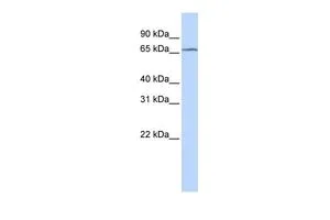
Anti-TRMT5 Antibody
HPA000943
ApplicationsWestern Blot, ImmunoCytoChemistry, ImmunoHistoChemistry
Product group Antibodies
ReactivityHuman
TargetTRMT5
Overview
- SupplierAtlas Antibodies
- Product NameAnti-TRMT5 Antibody
- Delivery Days Customer4
- ApplicationsWestern Blot, ImmunoCytoChemistry, ImmunoHistoChemistry
- CertificationResearch Use Only
- ClonalityPolyclonal
- ConjugateUnconjugated
- Gene ID57570
- Target nameTRMT5
- Target descriptiontRNA methyltransferase 5
- Target synonymsCOXPD26, KIAA1393, PNSED, TRM5, tRNA (guanine(37)-N(1))-methyltransferase, M1G-methyltransferase, TRM5 tRNA methyltransferase 5 homolog, tRNA (guanine(37)-N1)-methyltransferase, tRNA (guanine-N(1)-)-methyltransferase, tRNA [GM37] methyltransferase, tRNA-N1G37 methyltransferase
- HostRabbit
- IsotypeIgG
- Protein IDQ32P41
- Protein NametRNA (guanine(37)-N(1))-methyltransferase
- Scientific DescriptionRecombinant Protein Epitope Signature Tag (PrEST) antigen sequence
- ReactivityHuman
- Storage Instruction-20°C,2°C to 8°C
- UNSPSC41116161
Datasheet
MSDS
Related products
Product group Antibodies
TRMT5 AntibodyCSB-PA024541GA01HU
ApplicationsWestern Blot, ELISA
ReactivityHuman, Mouse, Rat
TargetTRMT5
- SizePrice

Product group Antibodies
Anti-TRMT5 Antibody Picoband(r)A11359-1-CARRIER-FREE
ApplicationsImmunoFluorescence, Western Blot, ELISA, ImmunoCytoChemistry
ReactivityHuman
TargetTRMT5
- SizePrice
Product group Antibodies

TRMT5 AntibodyLS-C752218
ApplicationsWestern Blot, ELISA
ReactivityHuman
TargetTRMT5
- SizePrice
Product group Antibodies
TRMT5 antibody, InternalGTX46177
ApplicationsWestern Blot
ReactivityHuman
TargetTRMT5
- SizePrice