


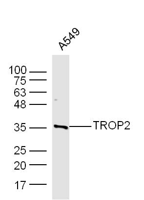
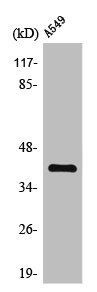
Figure 1. Western blot analysis of TROP2/TACSTD2 using anti-TROP2/TACSTD2 antibody (A03569-2). Electrophoresis was performed on a 5-20% SDS-PAGE gel at 70V (Stacking gel) / 90V (Resolving gel) for 2-3 hours. The sample well of each lane was loaded with 30 ug of sample under reducing conditions. Lane 1: human T-47D whole cell lysates, Lane 2: rat pancreas tissue lysates, Lane 3: mouse pancreas tissue lysates, Lane 4: mouse NIH/3T3 whole cell lysates. After electrophoresis, proteins were transferred to a nitrocellulose membrane at 150 mA for 50-90 minutes. Blocked the membrane with 5% non-fat milk/TBS for 1.5 hour at RT. The membrane was incubated with rabbit anti-TROP2/TACSTD2 antigen affinity purified polyclonal antibody (Catalog # A03569-2) at 0.5 microg/mL overnight at 4°C, then washed with TBS-0.1%Tween 3 times with 5 minutes each and probed with a goat anti-rabbit IgG-HRP secondary antibody at a dilution of 1:5000 for 1.5 hour at RT. The signal is developed using an Enhanced Chemiluminescent detection (ECL) kit (Catalog # EK1002) with Tanon 5200 system. A specific band was detected for TROP2/TACSTD2 at approximately 45-65 kDa. The expected band size for TROP2/TACSTD2 is at 36 kDa.
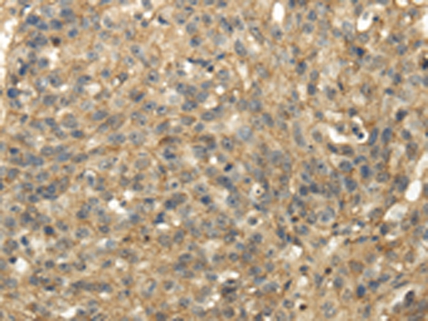
. TROP2/TACSTD2 was detected in an immunocytochemical section of PC-3 cells. Enzyme antigen retrieval was performed using IHC enzyme antigen retrieval reagent (AR0022) for 15 mins. The cells were blocked with 10% goat serum. And then incubated with 5 microg/mL rabbit anti-TROP2/TACSTD2 Antibody (A03569-2) overnight at 4°C. DyLight®488 Conjugated Goat Anti-Rabbit IgG (BA1127) was used as secondary antibody at 1:100 dilution and incubated for 30 minutes at 37°C. The section was counterstained with DAPI. Visualize using a fluorescence microscope and filter sets appropriate for the label used.")
. Overlay histogram showing RT4 cells stained with A03569-2 (Blue line). To facilitate intracellular staining, cells were fixed with 4% paraformaldehyde and permeabilized with permeabilization buffer. The cells were blocked with 10% normal goat serum. And then incubated with rabbit anti-TROP2/TACSTD2 Antibody (A03569-2, 1 microg/1x106 cells) for 30 min at 20°C. DyLight®488 conjugated goat anti-rabbit IgG (BA1127, 5-10 microg/1x106 cells) was used as secondary antibody for 30 minutes at 20°C. Isotype control antibody (Green line) was rabbit IgG (1 microg/1x106) used under the same conditions. Unlabelled sample without incubation with primary antibody and secondary antibody (Red line) was used as a blank control.")
Figure 1. Western blot analysis of TROP2/TACSTD2 using anti-TROP2/TACSTD2 antibody (A03569-2). Electrophoresis was performed on a 5-20% SDS-PAGE gel at 70V (Stacking gel) / 90V (Resolving gel) for 2-3 hours. The sample well of each lane was loaded with 30 ug of sample under reducing conditions. Lane 1: human T-47D whole cell lysates, Lane 2: rat pancreas tissue lysates, Lane 3: mouse pancreas tissue lysates, Lane 4: mouse NIH/3T3 whole cell lysates. After electrophoresis, proteins were transferred to a nitrocellulose membrane at 150 mA for 50-90 minutes. Blocked the membrane with 5% non-fat milk/TBS for 1.5 hour at RT. The membrane was incubated with rabbit anti-TROP2/TACSTD2 antigen affinity purified polyclonal antibody (Catalog # A03569-2) at 0.5 microg/mL overnight at 4°C, then washed with TBS-0.1%Tween 3 times with 5 minutes each and probed with a goat anti-rabbit IgG-HRP secondary antibody at a dilution of 1:5000 for 1.5 hour at RT. The signal is developed using an Enhanced Chemiluminescent detection (ECL) kit (Catalog # EK1002) with Tanon 5200 system. A specific band was detected for TROP2/TACSTD2 at approximately 45-65 kDa. The expected band size for TROP2/TACSTD2 is at 36 kDa.
Anti-TROP2/TACSTD2 Antibody Picoband(r)
A03569-2-CARRIER-FREE
ApplicationsFlow Cytometry, ImmunoFluorescence, Western Blot, ImmunoCytoChemistry
Product group Antibodies
ReactivityHuman, Mouse, Rat
TargetTACSTD2
Overview
- SupplierBoster Bio
- Product NameAnti-TROP2/TACSTD2 Antibody Picoband(r)
- Delivery Days Customer9
- ApplicationsFlow Cytometry, ImmunoFluorescence, Western Blot, ImmunoCytoChemistry
- CertificationResearch Use Only
- ClonalityPolyclonal
- Concentration500 ug/ml
- Gene ID4070
- Target nameTACSTD2
- Target descriptiontumor associated calcium signal transducer 2
- Target synonymsEGP-1, EGP1, GA733-1, GA7331, GP50, M1S1, TROP2, tumor-associated calcium signal transducer 2, 40kD glycoprotein, identified by monoclonal antibody GA733, cell surface glycoprotein TROP2, cell surface glycoprotein Trop-2, epithelial glycoprotein-1, gastrointestinal tumor-associated antigen GA7331, membrane component, chromosome 1, surface marker 1, pancreatic carcinoma marker protein GA733-1, pancreatic carcinoma marker protein GA7331, trophoblast cell surface antigen 2
- HostRabbit
- IsotypeIgG
- Protein IDP09758
- Protein NameTumor-associated calcium signal transducer 2
- Scientific DescriptionBoster Bio Anti-TROP2/TACSTD2 Antibody Picoband® catalog # A03569-2. Tested in Flow Cytometry, IF, ICC, WB applications. This antibody reacts with Human, Mouse, Rat. The brand Picoband indicates this is a premium antibody that guarantees superior quality, high affinity, and strong signals with minimal background in Western blot applications. Only our best-performing antibodies are designated as Picoband, ensuring unmatched performance.
- ReactivityHuman, Mouse, Rat
- Storage Instruction-20°C,2°C to 8°C
- UNSPSC12352203
Related products
Product group Antibodies
Anti-TACSTD2 AntibodyA45233
ApplicationsImmunoHistoChemistry
ReactivityHuman
- SizePrice
Product group Antibodies

Anti-TROP2 [hRS7 (Sacituzumab)]AB03992-10.17
ApplicationsFlow Cytometry, ImmunoHistoChemistry, Other Application
ReactivityHuman
TargetTACSTD2
- SizePrice
Product group Antibodies
References
TROP2 Polyclonal AntibodyBS-6198R
ApplicationsFlow Cytometry, ImmunoFluorescence, Western Blot, ELISA, ImmunoCytoChemistry, ImmunoHistoChemistry, ImmunoHistoChemistry Frozen, ImmunoHistoChemistry Paraffin
ReactivityHuman, Mouse, Rabbit, Rat
TargetTACSTD2
- SizePrice
Product group Antibodies
TACSTD2 AntibodyCSB-PA004333
ApplicationsWestern Blot, ELISA, ImmunoHistoChemistry
ReactivityHuman, Mouse, Rat
TargetTACSTD2
- SizePrice
Product group Antibodies

Goat anti-TROP2EB08839
ApplicationsWestern Blot, ELISA, ImmunoHistoChemistry
ReactivityBovine, Canine, Human, Mouse, Porcine, Rat
TargetTACSTD2
- SizePrice
Product group Antibodies
ApplicationsImmunoPrecipitation, Western Blot, ImmunoCytoChemistry, ImmunoHistoChemistry
TargetTACSTD2
- SizePrice
![IHC-P analysis of human renal cortex tissue using GTX04443 TACSTD2 antibody [MSVA-733R] HistoMAX?. Kidney cortex In the kidney TACSTD2 immunostaining is strong in distal tubuli and collecting ducts but weaker in the parietal layer of the Bowman capsule.](https://www.genetex.com/upload/website/prouct_img/normal/GTX04443/GTX04443_20230728_IHC-P_114_23072722_413.webp)
Product group Antibodies
ApplicationsImmunoHistoChemistry, ImmunoHistoChemistry Paraffin
ReactivityHuman
TargetTACSTD2
- SizePrice
Product group Antibodies

TROP2 / TACSTD2 AntibodyLS-C486602
ApplicationsFlow Cytometry, ImmunoFluorescence, ELISA, ImmunoCytoChemistry
ReactivityHuman
TargetTACSTD2
- SizePrice

Product group Antibodies
Anti-TACSTD2 AntibodyHPA043104
ApplicationsWestern Blot
ReactivityHuman
TargetTACSTD2
- SizePrice