


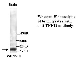
Figure 1. Western blot analysis of TNNI2 using anti-TNNI2 antibody (A07355-2). Electrophoresis was performed on a 5-20% SDS-PAGE gel at 70V (Stacking gel) / 90V (Resolving gel) for 2-3 hours. The sample well of each lane was loaded with 50ug of sample under reducing conditions. Lane 1: rat skeletal muscle tissue lysates, Lane 2: mouse skeletal muscle tissue lysates, After Electrophoresis, proteins were transferred to a Nitrocellulose membrane at 150mA for 50-90 minutes. Blocked the membrane with 5% Non-fat Milk/ TBS for 1.5 hour at RT. The membrane was incubated with rabbit anti-TNNI2 antigen affinity purified polyclonal antibody (Catalog # A07355-2) at 0.5 microg/mL overnight at 4°C, then washed with TBS-0.1%Tween 3 times with 5 minutes each and probed with a goat anti-rabbit IgG-HRP secondary antibody at a dilution of 1:10000 for 1.5 hour at RT. The signal is developed using an Enhanced Chemiluminescent detection (ECL) kit (Catalog # EK1002) with Tanon 5200 system. A specific band was detected for TNNI2 at approximately 24KD. The expected band size for TNNI2 is at 21KD.
. TNNI2 was detected in paraffin-embedded section of human skeletal muscle tissues. Heat mediated antigen retrieval was performed in citrate buffer (pH6, epitope retrieval solution) for 20 mins. The tissue section was blocked with 10% goat serum. The tissue section was then incubated with 1microg/ml rabbit anti-TNNI2 Antibody (A07355-2) overnight at 4°C. Biotinylated goat anti-rabbit IgG was used as secondary antibody and incubated for 30 minutes at 37°C. The tissue section was developed using Strepavidin-Biotin-Complex (SABC)(Catalog # SA1022) with DAB as the chromogen.")
. TNNI2 was detected in paraffin-embedded section of mouse skeletal muscle tissues. Heat mediated antigen retrieval was performed in citrate buffer (pH6, epitope retrieval solution) for 20 mins. The tissue section was blocked with 10% goat serum. The tissue section was then incubated with 1microg/ml rabbit anti-TNNI2 Antibody (A07355-2) overnight at 4°C. Biotinylated goat anti-rabbit IgG was used as secondary antibody and incubated for 30 minutes at 37°C. The tissue section was developed using Strepavidin-Biotin-Complex (SABC)(Catalog # SA1022) with DAB as the chromogen.")
. TNNI2 was detected in paraffin-embedded section of rat skeletal muscle tissues. Heat mediated antigen retrieval was performed in citrate buffer (pH6, epitope retrieval solution) for 20 mins. The tissue section was blocked with 10% goat serum. The tissue section was then incubated with 1microg/ml rabbit anti-TNNI2 Antibody (A07355-2) overnight at 4°C. Biotinylated goat anti-rabbit IgG was used as secondary antibody and incubated for 30 minutes at 37°C. The tissue section was developed using Strepavidin-Biotin-Complex (SABC)(Catalog # SA1022) with DAB as the chromogen.")
. TNNI2 was detected in a paraffin-embedded section of mouse skeletal muscle tissue. Heat mediated antigen retrieval was performed in EDTA buffer (pH 8.0, epitope retrieval solution). The tissue section was blocked with 10% goat serum. The tissue section was then incubated with 5 microg/mL rabbit anti-TNNI2 Antibody (A07355-2) overnight at 4°C. Biotin conjugated goat anti-rabbit IgG (BA1003) was used as secondary antibody and incubated for 30 minutes at 37°C. The tissue section was developed using Cy3 Conjugated Avidin (BA1037). The section was counterstained with DAPI. Visualize using a fluorescence microscope and filter sets appropriate for the label used.")
. TNNI2 was detected in a paraffin-embedded section of rat skeletal muscle tissue. Heat mediated antigen retrieval was performed in EDTA buffer (pH 8.0, epitope retrieval solution). The tissue section was blocked with 10% goat serum. The tissue section was then incubated with 5 microg/mL rabbit anti-TNNI2 Antibody (A07355-2) overnight at 4°C. Biotin conjugated goat anti-rabbit IgG (BA1003) was used as secondary antibody and incubated for 30 minutes at 37°C. The tissue section was developed using Cy3 Conjugated Avidin (BA1037). The section was counterstained with DAPI. Visualize using a fluorescence microscope and filter sets appropriate for the label used.")
Figure 1. Western blot analysis of TNNI2 using anti-TNNI2 antibody (A07355-2). Electrophoresis was performed on a 5-20% SDS-PAGE gel at 70V (Stacking gel) / 90V (Resolving gel) for 2-3 hours. The sample well of each lane was loaded with 50ug of sample under reducing conditions. Lane 1: rat skeletal muscle tissue lysates, Lane 2: mouse skeletal muscle tissue lysates, After Electrophoresis, proteins were transferred to a Nitrocellulose membrane at 150mA for 50-90 minutes. Blocked the membrane with 5% Non-fat Milk/ TBS for 1.5 hour at RT. The membrane was incubated with rabbit anti-TNNI2 antigen affinity purified polyclonal antibody (Catalog # A07355-2) at 0.5 microg/mL overnight at 4°C, then washed with TBS-0.1%Tween 3 times with 5 minutes each and probed with a goat anti-rabbit IgG-HRP secondary antibody at a dilution of 1:10000 for 1.5 hour at RT. The signal is developed using an Enhanced Chemiluminescent detection (ECL) kit (Catalog # EK1002) with Tanon 5200 system. A specific band was detected for TNNI2 at approximately 24KD. The expected band size for TNNI2 is at 21KD.
Anti-Troponin I fast skeletal muscle/TNNI2 Antibody Picoband(r)
A07355-2-CARRIER-FREE
ApplicationsImmunoFluorescence, Western Blot, ELISA, ImmunoHistoChemistry
Product group Antibodies
ReactivityHuman, Mouse, Rat
TargetTNNI2
Overview
- SupplierBoster Bio
- Product NameAnti-Troponin I fast skeletal muscle/TNNI2 Antibody Picoband(r)
- Delivery Days Customer9
- ApplicationsImmunoFluorescence, Western Blot, ELISA, ImmunoHistoChemistry
- CertificationResearch Use Only
- ClonalityPolyclonal
- Concentration500 ug/ml
- Gene ID7136
- Target nameTNNI2
- Target descriptiontroponin I2, fast skeletal type
- Target synonymsAMCD2B, DA2B, DA2B1, FSSV, fsTnI, troponin I, fast skeletal muscle, troponin I fast twitch 2, troponin I type 2 (skeletal, fast), troponin I, skeletal, fast
- HostRabbit
- IsotypeIgG
- Protein IDP48788
- Protein NameTroponin I, fast skeletal muscle
- Scientific DescriptionBoster Bio Anti-Troponin I fast skeletal muscle/TNNI2 Antibody Picoband® catalog # A07355-2. Tested in ELISA, IF, IHC, WB applications. This antibody reacts with Human, Mouse, Rat. The brand Picoband indicates this is a premium antibody that guarantees superior quality, high affinity, and strong signals with minimal background in Western blot applications. Only our best-performing antibodies are designated as Picoband, ensuring unmatched performance.
- ReactivityHuman, Mouse, Rat
- Storage Instruction-20°C,2°C to 8°C
- UNSPSC12352203
Related products
Product group Antibodies
Anti-TNNI2 AntibodyA49290
ApplicationsWestern Blot, ELISA
ReactivityHuman, Mouse, Rat
- SizePrice
Product group Antibodies
Anti-TNNI2 Antibody144-07937
ApplicationsWestern Blot
ReactivityHuman, Mouse, Rat
TargetTNNI2
- SizePrice
Product group Antibodies
TNNI2 Recombinant AntibodyBSM-62039R
ApplicationsImmunoPrecipitation, Western Blot
ReactivityHuman, Mouse, Rat
TargetTNNI2
- SizePrice
Product group Antibodies

Goat anti-TNNI2EB12036
ApplicationsWestern Blot, ELISA
ReactivityCanine, Human, Mouse, Rat
TargetTNNI2
- SizePrice
Product group Antibodies
TNNI2 AntibodyCSB-PA024012ESR2HU
ApplicationsImmunoPrecipitation, Western Blot, ELISA, ImmunoHistoChemistry
ReactivityHuman, Mouse
TargetTNNI2
- SizePrice
Product group Antibodies
ApplicationsImmunoPrecipitation, Western Blot, ImmunoCytoChemistry, ImmunoHistoChemistry
ReactivityMouse, Porcine, Rat
TargetTNNI2
- SizePrice
Product group Antibodies

TNNI2 AntibodyLS-C409484
ApplicationsWestern Blot, ImmunoHistoChemistry
ReactivityHuman, Mouse, Rat
TargetTNNI2
- SizePrice
Product group Antibodies
ApplicationsWestern Blot, Purification/Extraction/Isolation
ReactivityHuman
TargetTNNI2
- SizePrice

Product group Antibodies
Anti-TNNI2 AntibodyHPA055938
ApplicationsWestern Blot, ImmunoCytoChemistry, ImmunoHistoChemistry
ReactivityHuman
TargetTNNI2
- SizePrice