


Immunohistochemical staining of human cerebral cortex shows moderate cytoplasmic positivity in neuronal cells.

Immunohistochemical staining of human cerebral cortex shows moderate cytoplasmic positivity in neuronal cells.
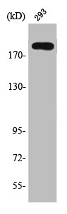
Anti-TSC2 Antibody
HPA030409
ApplicationsWestern Blot, ImmunoCytoChemistry, ImmunoHistoChemistry
Product group Antibodies
ReactivityHuman
TargetTSC2
Overview
- SupplierAtlas Antibodies
- Product NameAnti-TSC2 Antibody
- Delivery Days Customer4
- ApplicationsWestern Blot, ImmunoCytoChemistry, ImmunoHistoChemistry
- CertificationResearch Use Only
- ClonalityPolyclonal
- ConjugateUnconjugated
- Gene ID7249
- Target nameTSC2
- Target descriptionTSC complex subunit 2
- Target synonymsLAM, PPP1R160, TSC4, tuberin, protein phosphatase 1, regulatory subunit 160, tuberous sclerosis 2 protein
- HostRabbit
- IsotypeIgG
- Protein IDP49815
- Protein NameTuberin
- Scientific DescriptionRecombinant Protein Epitope Signature Tag (PrEST) antigen sequence
- ReactivityHuman
- Storage Instruction-20°C,2°C to 8°C
- UNSPSC41116161
Datasheet
MSDS
Related products
Product group Antibodies
TSC2 AntibodyCSB-PA004343
ApplicationsImmunoFluorescence, Western Blot, ELISA, ImmunoHistoChemistry
ReactivityHuman, Mouse, Rat
TargetTSC2
- SizePrice
Product group Antibodies

Anti-TSC2 [RAB-C431]Ab01910-1.1
ApplicationsImmunoPrecipitation
ReactivityHuman
TargetTSC2
- SizePrice
Product group Antibodies
ApplicationsWestern Blot
ReactivityHuman, Mouse, Rat
- SizePrice

Product group Antibodies
Anti-TSC2 AntibodyHPA049679
ApplicationsImmunoCytoChemistry
ReactivityHuman
TargetTSC2
- SizePrice
Product group Antibodies

Tuberin / TSC2 AntibodyLS-C496666
ApplicationsImmunoFluorescence, Western Blot, ImmunoHistoChemistry
ReactivityHuman
TargetTSC2
- SizePrice
Product group Antibodies
TSC2 Polyclonal AntibodyCAC14612
ApplicationsImmunoFluorescence, Western Blot, ELISA
ReactivityMouse
TargetTSC2
- SizePrice

Product group Antibodies
Anti-Tuberin/TSC2 Antibody Picoband(r)PB9121-CARRIER-FREE
ApplicationsWestern Blot, ImmunoHistoChemistry
ReactivityHuman, Mouse, Rat
TargetTSC2
- SizePrice