


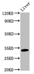
Anti-TST Antibody
A12986
ApplicationsImmunoPrecipitation, Western Blot
Product group Antibodies
ReactivityHuman, Mouse, Rat
Overview
- SupplierAntibodies.com
- Product NameAnti-TST Antibody
- Delivery Days Customer7
- ApplicationsImmunoPrecipitation, Western Blot
- CertificationResearch Use Only
- ClonalityPolyclonal
- ConjugateUnconjugated
- HostRabbit
- IsotypeIgG
- Scientific DescriptionRabbit polyclonal antibody to TST.
- ReactivityHuman, Mouse, Rat
- UNSPSC12352203
Related products
Product group Antibodies
TST Polyclonal AntibodyCAC13349
ApplicationsWestern Blot, ELISA, ImmunoHistoChemistry
ReactivityRat
TargetTST
- SizePrice
Product group Antibodies
Anti-TST Antibody144-10542
ApplicationsImmunoPrecipitation, Western Blot
ReactivityHuman, Mouse, Rat
TargetTST
- SizePrice
Product group Antibodies
TST antibodyGTX106257
ApplicationsWestern Blot, ImmunoHistoChemistry, ImmunoHistoChemistry Paraffin
ReactivityHuman, Mouse, Rat
TargetTST
- SizePrice
Product group Antibodies
TST AntibodyCSB-PA620980LA01HU
ApplicationsWestern Blot, ELISA, ImmunoHistoChemistry
ReactivityHuman, Rat
TargetTST
- SizePrice
Product group Antibodies

Rhodanese / TST AntibodyLS-C497253
ApplicationsWestern Blot
ReactivityHuman, Mouse, Rat
TargetTST
- SizePrice

Product group Antibodies
Anti-TST AntibodyHPA003044
ApplicationsImmunoCytoChemistry, ImmunoHistoChemistry
ReactivityHuman
TargetTST
- SizePrice

Product group Antibodies
Anti-TST Antibody Picoband(r)A00965-1-CARRIER-FREE
ApplicationsImmunoFluorescence, Western Blot, ELISA, ImmunoCytoChemistry, ImmunoHistoChemistry
ReactivityHuman, Mouse, Rat
TargetTST
- SizePrice