

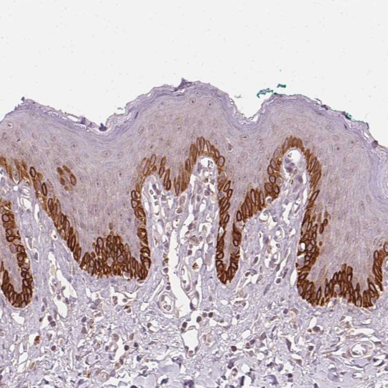
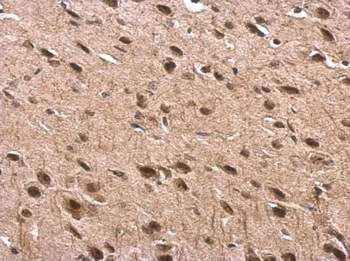
Immunohistochemical staining of human cerebellum shows strong cytoplasmic positivity in Purkinje cells.

Immunohistochemical staining of human cerebellum shows strong cytoplasmic positivity in Purkinje cells.
Anti-TUB Antibody
HPA017997
ApplicationsImmunoCytoChemistry, ImmunoHistoChemistry
Product group Antibodies
ReactivityHuman
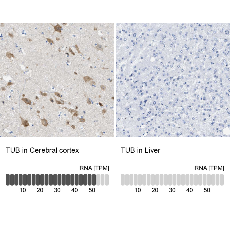
TargetTUB
Overview
- SupplierAtlas Antibodies
- Product NameAnti-TUB Antibody
- Delivery Days Customer4
- ApplicationsImmunoCytoChemistry, ImmunoHistoChemistry
- CertificationResearch Use Only
- ClonalityPolyclonal
- ConjugateUnconjugated
- Gene ID7275
- Target nameTUB
- Target descriptionTUB bipartite transcription factor
- Target synonymsRDOB, rd5, tubby protein homolog, tubby bipartite transcription factor, tubby homolog, tubby homologue
- HostRabbit
- IsotypeIgG
- Protein IDP50607
- Protein NameTubby protein homolog
- Scientific DescriptionRecombinant Protein Epitope Signature Tag (PrEST) antigen sequence
- ReactivityHuman
- Storage Instruction-20°C,2°C to 8°C
- UNSPSC41116161
Datasheet
MSDS
Related products
Product group Antibodies
Anti-TUB 1 AntibodyA306737
ApplicationsWestern Blot
ReactivityHuman, Rat
- SizePrice
Product group Antibodies
Anti-TUB 1 Antibody Picoband(r)A02917-1-CARRIER-FREE
ApplicationsFlow Cytometry, Western Blot
ReactivityHuman
TargetTUB
- SizePrice
Product group Antibodies

TUB / Tubby AntibodyLS-C662171
ApplicationsWestern Blot
ReactivityHuman
TargetTUB
- SizePrice
Product group Antibodies
TUB 1 Polyclonal AntibodyBS-11536R
ApplicationsImmunoFluorescence, Western Blot, ELISA, ImmunoCytoChemistry, ImmunoHistoChemistry, ImmunoHistoChemistry Frozen, ImmunoHistoChemistry Paraffin
ReactivityBovine, Chicken, Equine, Human, Mouse, Porcine, Rat
- SizePrice
Product group Antibodies
TUB AntibodyCSB-PA025305GA01HU
ApplicationsWestern Blot, ELISA, ImmunoHistoChemistry
ReactivityHuman, Mouse, Rat
TargetTUB
- SizePrice
Product group Antibodies
Anti-TUB AntibodyHPA049019
ApplicationsImmunoHistoChemistry
ReactivityHuman
TargetTUB
- SizePrice
Product group Antibodies
Tub antibodyGTX118348
ApplicationsImmunoFluorescence, Western Blot, ImmunoCytoChemistry, ImmunoHistoChemistry, ImmunoHistoChemistry Paraffin
ReactivityHuman, Mouse, Rat
TargetTUB
- SizePrice