


Immunohistochemical staining of human cerebral cortex shows moderate cytoplasmic positivity in neuropil.
 shows similar pattern to independent antibody HPA039247 (B).")
Immunohistochemical staining of human cerebral cortex shows moderate cytoplasmic positivity in neuropil.
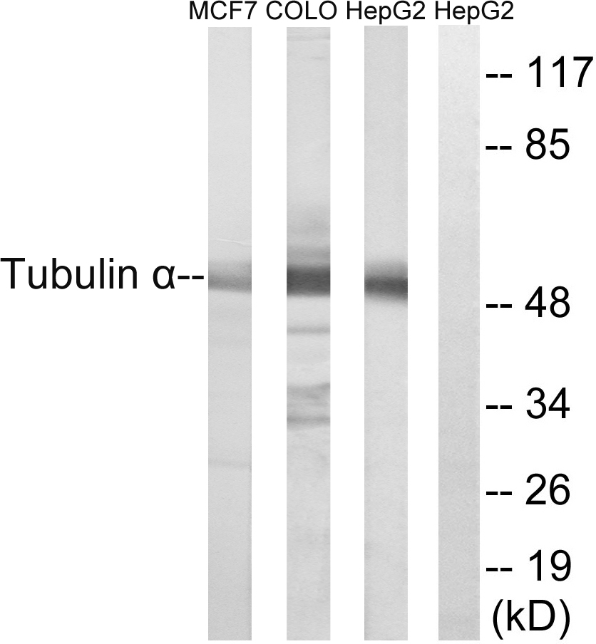
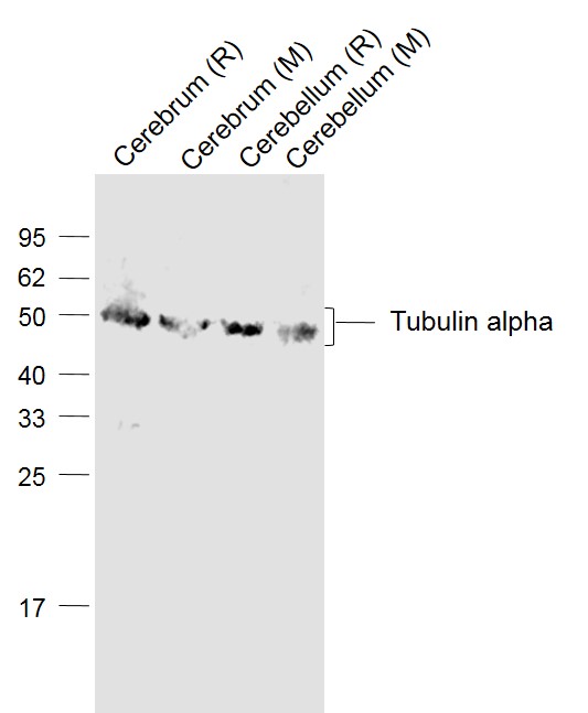
Anti-TUBA1A Antibody
HPA043684
ApplicationsWestern Blot, ImmunoCytoChemistry, ImmunoHistoChemistry
Product group Antibodies
ReactivityHuman, Mouse, Rat
TargetTUBA1A
Overview
- SupplierAtlas Antibodies
- Product NameAnti-TUBA1A Antibody
- Delivery Days Customer4
- ApplicationsWestern Blot, ImmunoCytoChemistry, ImmunoHistoChemistry
- CertificationResearch Use Only
- ClonalityPolyclonal
- ConjugateUnconjugated
- Gene ID7846
- Target nameTUBA1A
- Target descriptiontubulin alpha 1a
- Target synonymsB-ALPHA-1, LIS3, TUBA3, tubulin alpha-1A chain, hum-a-tub1, hum-a-tub2, tubulin B-alpha-1, tubulin alpha-3 chain, tubulin, alpha, brain-specific
- HostRabbit
- IsotypeIgG
- Protein IDQ71U36
- Protein NameTubulin alpha-1A chain
- Scientific DescriptionRecombinant Protein Epitope Signature Tag (PrEST) antigen sequence
- ReactivityHuman, Mouse, Rat
- Storage Instruction-20°C,2°C to 8°C
- UNSPSC41116161
Datasheet
MSDS
Related products
Product group Antibodies
ApplicationsImmunoFluorescence, Western Blot, ELISA, ImmunoHistoChemistry
ReactivityHuman, Mouse, Rat
- SizePrice
Product group Antibodies

Anti-Alpha-Tubulin [F2C]Ab00403-1.1
ApplicationsImmunoFluorescence, Western Blot, ELISA
ReactivityHuman
TargetTUBA1A
- SizePrice
Product group Antibodies
Anti-Tubulin alpha Antibody102-25843
ApplicationsImmunoFluorescence, Western Blot, ImmunoHistoChemistry
TargetTUBA1A
- SizePrice

Product group Antibodies
Anti-Tubulin alpha Antibody Picoband(r)A03989-1-CARRIER-FREE
ApplicationsFlow Cytometry, ImmunoFluorescence, Western Blot, ELISA, ImmunoCytoChemistry, ImmunoHistoChemistry
ReactivityHuman, Mouse, Rat
TargetTUBA1A
- SizePrice
Product group Antibodies
Tubulin alpha Polyclonal AntibodyBS-20496R
ApplicationsImmunoFluorescence, Western Blot, ELISA, ImmunoCytoChemistry, ImmunoHistoChemistry, ImmunoHistoChemistry Frozen, ImmunoHistoChemistry Paraffin
ReactivityHuman, Mouse, Rat
TargetTUBA1A
- SizePrice
Product group Antibodies
TUBA1A Monoclonal AntibodyCSB-MA000190
ApplicationsImmunoPrecipitation, Western Blot, ELISA
ReactivityHuman, Mouse, Rat
TargetTUBA1A
- SizePrice
Product group Antibodies
TUBA1A Monoclonal AntibodyCAC12980
ApplicationsFlow Cytometry, ImmunoFluorescence, ImmunoPrecipitation, Western Blot, ELISA, ImmunoHistoChemistry
ReactivityMouse, Rabbit, Rat
- SizePrice
Product group Antibodies

TUBA1A / Tubulin Alpha 1a Antibody (HRP)LS-C377875
ApplicationsELISA, ImmunoHistoChemistry
ReactivityHuman
TargetTUBA1A
- SizePrice